University Biology — Year 3 · Bachelor Year 3
10Cell Cycle Control and Programmed Cell Death
About a hundred billion cells of your body will die today, on purpose, and a hundred billion will be born to replace them; the lining of your gut is renewed every five days, and your red cells every four months. A tadpole loses its tail without bleeding; the webbing between a human embryo’s fingers vanishes in the eighth week; half the neurons ever made in a brain are killed before birth. Every one of these deaths is executed by the cell itself, on a programme, and every one of the births is licensed by a control system that decides, at two or three points in the cycle, whether the cell may proceed. The two programmes — division and suicide — are the subject of this chapter. They were worked out in yeast, frog eggs, sea urchins and a worm with exactly somatic cells of which exactly die, and they turned out to be the same in humans, where their failures are cancer on one side and degeneration on the other.
10.1 The engine of the cycle
Definition 10.1 (Cyclins and cyclin-dependent kinases)
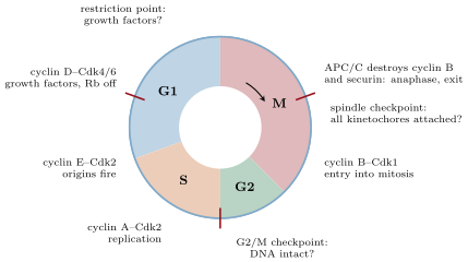
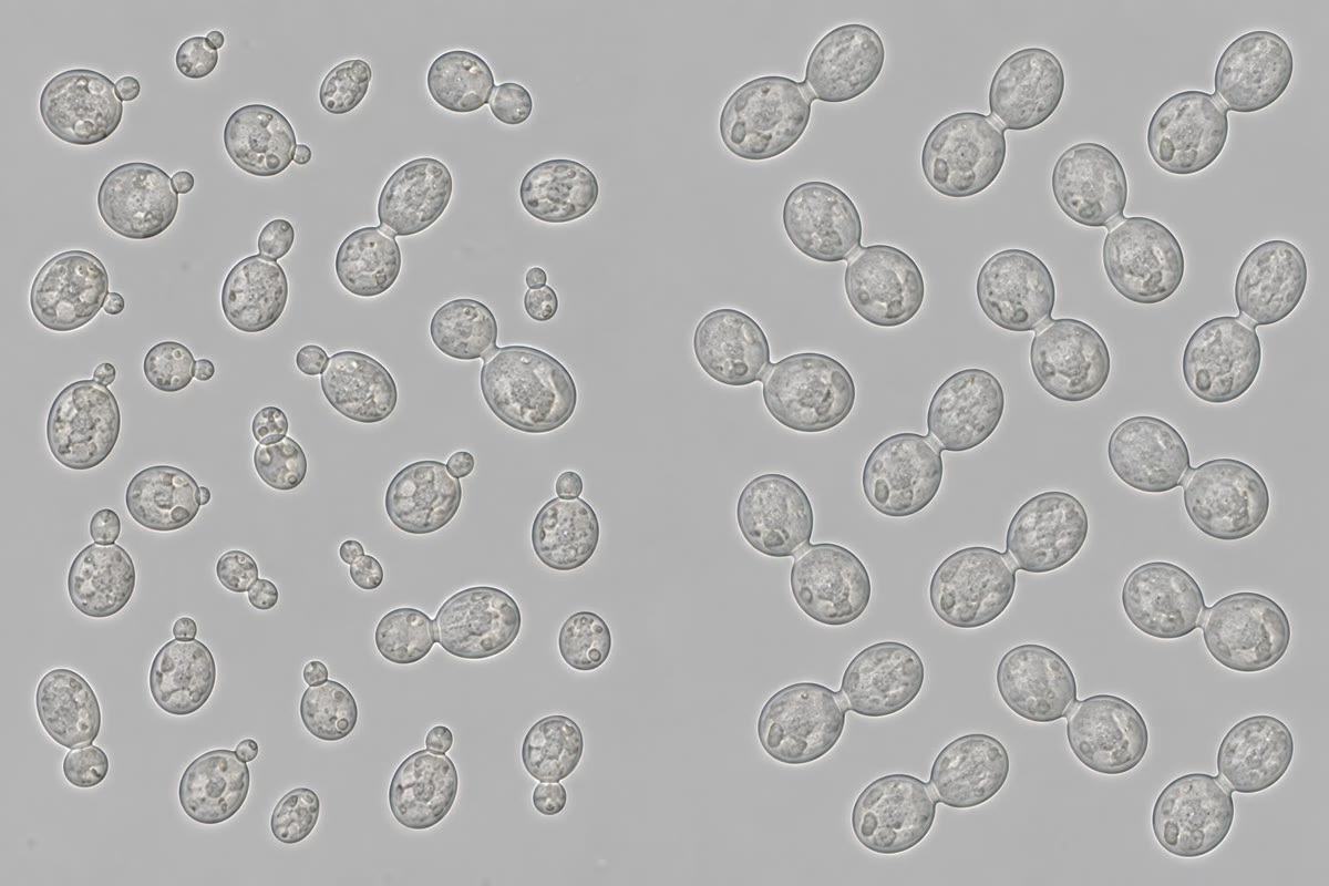
The cell cycle — G1, S (replication), G2, M (mitosis) — is driven by a family of protein kinases, the cyclin-dependent kinases (CDKs), whose catalytic subunits are present throughout the cycle but active only when bound to a cyclin, a regulatory subunit whose concentration rises and falls in a fixed order: cyclin D with Cdk4/6 in G1 in response to growth factors, cyclin E with Cdk2 at the G1/S transition, cyclin A with Cdk2 through S, cyclin B with Cdk1 at the entry into mitosis. Each cyclin–CDK phosphorylates the proteins of its phase — replication origins, lamins, condensins, the enzymes of spindle assembly — and each is switched off by the destruction of its cyclin: ubiquitin ligases (SCF in G1/S, the anaphase-promoting complex, APC/C, in mitosis) tag the cyclins for the proteasome. The activity of Cdk1 is further gated by an inhibitory phosphorylation put on by the kinase Wee1 and removed by the phosphatase Cdc25, and by small inhibitor proteins (p21, p27, p16) that bind the complexes. The cycle is thus an ordered sequence of kinase waves, each wave ending in the proteolysis of what produced it.
Evidence. Three lines converged. Hartwell (1970s) isolated temperature-sensitive cdc mutants of budding yeast, each arrested at one stage with a characteristic bud size, and defined Start, the point in G1 after which a cell is committed to a cycle. Nurse (1980s) found in fission yeast that cdc2 controlled the timing of mitosis and that the human gene, put into the yeast, rescued the mutant: the kinase was Cdk1, conserved from yeast to man. Hunt (1983) found in sea urchin eggs a protein that accumulated through each cycle and was destroyed abruptly at every division, and called it cyclin. The frog egg had meanwhile yielded a “maturation-promoting factor” that drove any nucleus into mitosis; it was cyclin B bound to Cdk1. Rao and Johnson (1970) had shown by fusing cells that an S-phase cell drives a G1 nucleus into replication, while a G2 nucleus waits — the cytoplasm carries the signal, and a replicated nucleus cannot be made to replicate again. ∎
Theorem 10.2 (A switch and a delayed brake make an oscillator)
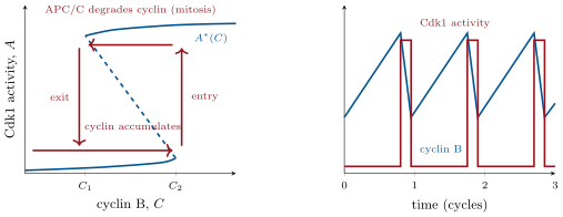
Let be the activity of cyclin B–Cdk1 and the amount of cyclin B. Suppose (i) for a fixed , relaxes quickly to a steady state that depends on through a positive feedback (active Cdk1 activates its activator Cdc25 and inhibits its inhibitor Wee1), so that the steady-state curve is S-shaped: for between two thresholds both a low and a high state exist, and the system stays on whichever branch it is on (hysteresis); (ii) changes slowly, synthesised at a constant rate and degraded at a rate through the APC/C, which active Cdk1 switches on after a delay. Then the system has no stable steady state and executes a relaxation oscillation: accumulates on the low branch until it passes , jumps to the high branch (mitotic entry), degradation outruns synthesis and falls until it passes , collapses to the low branch (mitotic exit), and the cycle repeats. The period is set mainly by the slow variable: about for the rise, plus the time to degrade from to .
Partial proof. A steady state of the pair would need , that is , at a point on the S-shaped curve. On the low branch is small, so for up to : cyclin rises, and the system is carried off the branch’s end. On the high branch is large, so for down to : cyclin falls, and the system is carried off that end. The only candidate steady state would lie on the middle, unstable branch, and a point there repels; hence there is no stable steady state, and the trajectory, confined to the two stable branches and the jumps between them, cycles. The time on the low branch is when is small, and the time on the high branch is the shorter time to degrade at rate . The existence of the S-shaped curve from the Cdc25/Wee1 feedback is admitted here; it is the same bistability as the self-activating gene of the Year 2 volume, with the fast variable now a kinase and the slow one its cyclin. ∎
Example 10.3 (Why the frog egg is a clock)
A fertilised frog egg divides twelve times in six hours with no growth, no transcription and no checkpoints, at intervals of : it is the oscillator of the theorem running bare, and an extract of its cytoplasm in a test tube goes on cycling, cyclin rising and falling, with nothing to divide. Adding a non-degradable cyclin B locks the extract in mitosis, since can never fall below ; blocking cyclin synthesis locks it in interphase. In a somatic cell the same engine is wrapped in the controls of the next section, which hold it at the thresholds until conditions are met, so that the period becomes a day rather than half an hour and can be indefinitely long.
10.2 Checkpoints
Definition 10.4 (Checkpoints)
A checkpoint is a control that halts the cycle at a transition until a condition is satisfied, by acting on the engine — inhibiting a CDK or an ubiquitin ligase. Three matter most. The restriction point in late G1 (Start in yeast): the cell proceeds only if growth factors, nutrients and size permit; beyond it the cycle completes without them. The G2/M checkpoint: damaged or unreplicated DNA, signalled by the ATM/ATR kinases of Chapter 3, keeps Cdc25 inhibited and so keeps Cdk1 phosphorylated and inactive. The spindle assembly checkpoint: a single kinetochore not attached to microtubules produces a diffusible inhibitor (Mad2 bound to Cdc20) that keeps the APC/C off, so that anaphase waits for the last chromosome. A fourth control has no waiting step but is equally strict: replication licensing. Origins are loaded with the MCM helicase only in G1, when CDK activity is low, and the loading factors are destroyed or inhibited once S phase begins, so that each origin fires once and only once per cycle — the reason a G2 nucleus in Rao and Johnson’s fusions could not be made to replicate.
Proposition 10.5 (The restriction point is a switch)
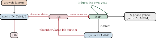
In early G1 the transcription factor E2F, which drives the genes of S phase (cyclin E, cyclin A, the replication enzymes), is held inactive by the retinoblastoma protein Rb. Growth factors induce cyclin D, and cyclin D–Cdk4/6 begins to phosphorylate Rb, releasing a little E2F; E2F then induces cyclin E, and cyclin E–Cdk2 phosphorylates Rb much more — a positive feedback that, once past a threshold, completes itself without further growth factor. The cell has crossed the restriction point; E2F also induces its own gene. Loss of Rb, or of the Cdk4/6 inhibitor p16, or amplification of cyclin D, removes the requirement for growth factor altogether, and each is common in cancer (Chapter 11); drugs that inhibit Cdk4/6 are now used against breast cancers that depend on this switch.
Proposition 10.6 (Anaphase is a proteolytic decision)
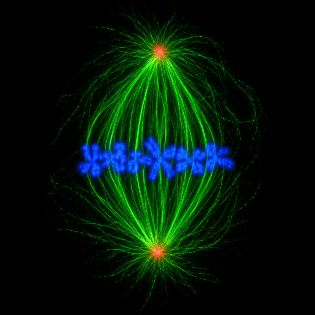
Sister chromatids are held together from S phase by rings of cohesin. At metaphase, when every kinetochore is attached and the spindle checkpoint is silenced, the APC/C with its activator Cdc20 ubiquitinates two proteins: cyclin B, whose loss inactivates Cdk1 and allows the cell to exit mitosis, and securin, whose loss frees the protease separase, which cuts cohesin. All sister pairs separate within seconds of each other, pulled to the poles by the spindle, and the cell divides by a ring of actin and myosin II assembled where the spindle midzone tells the cortex (through RhoA). The decision is irreversible because it is proteolytic: a cut cohesin cannot be reassembled, and the degraded cyclin must be resynthesised. Errors here — a chromosome pulled to the wrong pole, an anaphase begun before the last attachment — produce aneuploid daughters, the commonest chromosomal abnormality of tumours and of miscarriages.
10.3 Apoptosis
Definition 10.7 (Apoptosis)
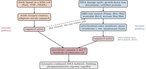
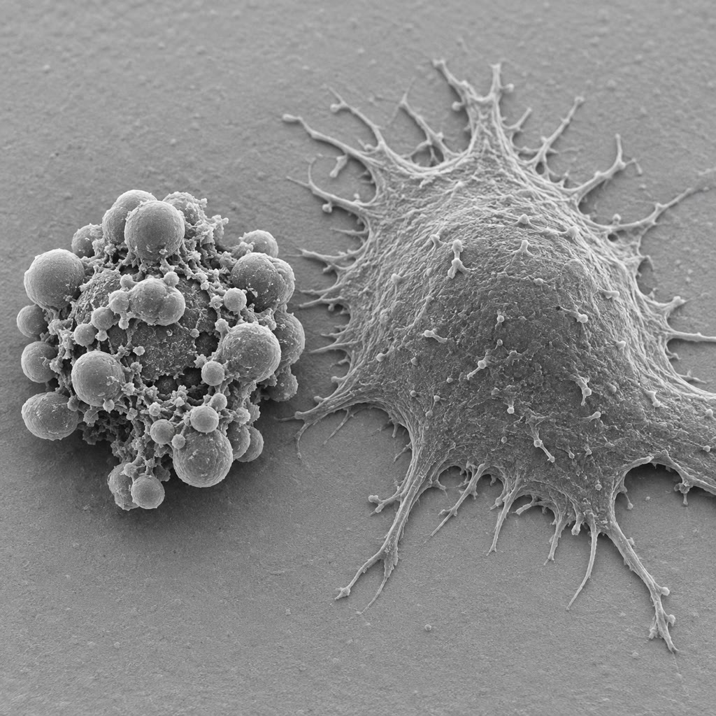
Apoptosis is programmed cell death by an intrinsic sequence: the cell shrinks and rounds up, its chromatin condenses and its DNA is cut between nucleosomes into a ladder of fragments, the nucleus breaks up, the membrane blebs into sealed vesicles, phosphatidylserine appears on the outer face of the membrane as an “eat me” signal, and the fragments are engulfed by neighbours or macrophages within an hour — without leakage, and therefore without inflammation. It is executed by caspases, cysteine proteases that cut after aspartate, made as inactive zymogens: initiator caspases (8 and 9) are activated by being brought together on a platform, and they cleave and activate the executioner caspases (3 and 7), which cut several hundred substrates — the lamins, the cytoskeleton, the inhibitor of the DNA-cutting nuclease, the enzyme that keeps phosphatidylserine inside. Necrosis, by contrast, is death by injury: the cell swells, bursts, spills its contents and provokes inflammation. Apoptosis was named and its morphology described by Kerr, Wyllie and Currie (1972); its genes were found in a worm.
Evidence. Every Caenorhabditis elegans hermaphrodite makes somatic cells, of which the same die, each at a fixed time and place. Horvitz and colleagues (1980s) isolated mutants in which they survived: ced-3 and ced-4 were needed for every death, and in their absence the cells lived and differentiated; ced-9 protected — its loss killed cells that should have lived, its over-activity saved cells that should have died; egl-1 was needed for particular deaths. CED-3 was a caspase, CED-4 its activating platform (Apaf-1 in humans), CED-9 a Bcl-2 protein, EGL-1 a BH3-only protein: the human pathway, gene for gene. The human BCL2 gene had itself been found at a chromosome translocation in follicular lymphoma, a cancer of cells that fail to die. ∎
Definition 10.8 (The two pathways)
The intrinsic (mitochondrial) pathway integrates the cell’s internal state. The Bcl-2 family has three classes: guardians (Bcl-2, Bcl-xL, Mcl-1) that sit on the mitochondrial outer membrane and keep it sealed; effectors (Bax, Bak) that, when activated, oligomerise into pores in that membrane; and BH3-only sensors (Bim, Bid, Puma, Noxa, Bad), each induced or activated by a particular insult — DNA damage through p53 (Puma, Noxa), growth-factor withdrawal (Bim, Bad), detachment, unfolded proteins — which bind the guardians and free or activate the effectors. When effectors win, the outer membrane is permeabilised, cytochrome c leaks into the cytosol, binds Apaf-1 and forms the apoptosome, which activates caspase 9. The extrinsic pathway starts outside: death receptors (Fas, TNF receptor, TRAIL receptors) bound by their ligands on a killer lymphocyte or a neighbour cluster, recruit adaptors and activate caspase 8, which cleaves caspase 3 directly and, through Bid, also opens the mitochondrial route. Both pathways converge on the executioner caspases, and inhibitor proteins (IAPs, FLIP) set the threshold along the way.
Proposition 10.9 (The point of no return)
The permeabilisation of the mitochondrial outer membrane is sudden — all the mitochondria of a cell open within about five minutes, after hours of deliberation — and complete: once cytochrome is in the cytosol, caspase activation follows in minutes and cannot be undone by removing the stimulus. Two features make it a switch. The Bax/Bak pore forms by oligomerisation, so its rate rises steeply with the amount of activated effector; and the guardians and sensors bind one another with high affinity, so that up to a threshold every sensor is neutralised and beyond it every extra sensor is free — a titration, like a buffer being exhausted. The cell therefore does not die gradually; it accumulates BH3-only proteins until a threshold is crossed, and then dies all at once. Drugs that mimic BH3 proteins (venetoclax, which occupies Bcl-2) push cells that are “primed” — already near the threshold, as many leukaemic cells are — over it, while sparing cells that are not.
Example 10.10 (Deaths that build a body)
The fingers are carved from a paddle by the apoptosis of the cells between them; in the duck the same cells live and the webbing stays. The tadpole’s tail is resorbed at metamorphosis on the signal of thyroid hormone. About half of the neurons produced in the vertebrate nervous system die before birth, those that fail to receive enough trophic factor from their targets — a competition that matches the number of neurons to the size of the field they serve. Ninety-five per cent of the T cells made in the thymus die there, selected away because they recognise nothing or recognise the self (Chapter 16). And every day the intestinal epithelium sheds its oldest cells at the villus tips, the skin its keratinocytes, the blood its aged neutrophils after a life of a day: apoptosis is the routine, and the tissue’s size is the difference between two large rates.
10.4 Other exits, and the failure to exit
Definition 10.11 (Regulated necrosis and senescence)
Cells have other programmed deaths, all inflammatory where apoptosis is silent. Necroptosis, triggered by death receptors when caspase-8 is blocked (as some viruses block it), assembles the kinase RIPK3 with MLKL, which punctures the plasma membrane. Pyroptosis, in infected macrophages, is executed by caspase-1 of the inflammasome (Chapter 15), which cleaves gasdermin into a pore-former and releases interleukin-1. Ferroptosis is death by iron-dependent lipid peroxidation. A cell can also stop dividing without dying: senescence is a permanent arrest, enforced by p16 and p21 after telomere erosion (Chapter 24), persistent DNA damage or oncogene activation, in which the cell stays metabolically active and secretes inflammatory factors. Senescence removes damaged cells from the dividing pool — a tumour suppressor — and accumulates with age, where its secretions contribute to the degeneration of tissues; clearing senescent cells in mice extends healthy life.
Remark 10.12 (Too little and too much)
The diseases of these programmes are of dose. Too little death: cancer, in which the apoptotic threshold is raised by Bcl-2 overexpression or p53 loss, so that cells with damaged genomes survive to accumulate more damage; autoimmunity, when lymphocytes that should have died in selection persist. Too much: the loss of T cells in HIV infection, the neurons of a stroke’s penumbra dying by apoptosis hours after the ischaemia (a window for treatment), the slow apoptosis of neurons in neurodegenerative disease, the cardiac cells lost after a heart attack. Cancer treatment is largely the deliberate induction of apoptosis in cells whose thresholds are lower than their neighbours’, and the drugs that target Bcl-2 and Cdk4/6 are the first designed from the maps of this chapter.
10.5 Exercises
Exercise 10.1 ★
List the four phases of the cycle, the cyclin–CDK pair that drives each transition, and the ubiquitin ligase that ends mitosis.
Solution
Solution of Exercise 10.1.
G1, S, G2, M. G1 progression and the restriction point: cyclin D–Cdk4/6; G1/S: cyclin E–Cdk2; S: cyclin A–Cdk2; G2/M: cyclin B–Cdk1. The anaphase-promoting complex (APC/C) destroys cyclin B and securin and ends mitosis.
Exercise 10.2 ★
What did each of Hartwell, Nurse and Hunt contribute to the discovery of the engine, and in what organism?
Solution
Solution of Exercise 10.2.
Hartwell: the cdc mutants of budding yeast, arrested at defined stages, and the concept of Start. Nurse: cdc2 of fission yeast as the kinase timing mitosis, and its human homologue (Cdk1) rescuing the yeast — conservation. Hunt: cyclin in sea urchin eggs, a protein synthesised through the cycle and destroyed at each division.
Exercise 10.3 ★
Name the three checkpoints, the condition each tests, and the molecular target each acts on.
Solution
Solution of Exercise 10.3.
Restriction point (late G1): growth factors, nutrients, size; acts on cyclin D–Cdk4/6 and Rb. G2/M: DNA intact and replicated; ATM/ATR inhibit Cdc25, keeping Cdk1 phosphorylated and inactive. Spindle assembly: every kinetochore attached; unattached kinetochores make Mad2–Cdc20 complexes that inhibit the APC/C.
Exercise 10.4 ★
Distinguish apoptosis from necrosis in five features, and name the class of enzyme that executes apoptosis.
Solution
Solution of Exercise 10.4.
Apoptosis: cell shrinks, chromatin condenses, DNA cut into a ladder, membrane blebs but stays sealed, phosphatidylserine exposed, corpse engulfed, no inflammation. Necrosis: cell swells, membrane ruptures, contents leak, random DNA degradation, inflammation. Executioners: caspases, cysteine proteases cutting after aspartate.
Exercise 10.5 ★★
In a frog egg extract cyclin B is synthesised at unit per minute; the mitotic threshold is units, the exit threshold , and in mitosis cyclin is degraded with a half-life of . Estimate the period of the oscillation and the fraction of the cycle spent in mitosis.
Solution
Solution of Exercise 10.5.
Rise from to at per minute: . Degradation from to with half-life : . Period about , mitosis of it.
Exercise 10.6 ★★
Predict the behaviour of a cell (a) expressing a non-degradable cyclin B, (b) lacking Wee1, (c) lacking Cdc25, (d) expressing a Cdk1 that cannot be phosphorylated by Wee1, in terms of the oscillator of Theorem 10.2.
Solution
Solution of Exercise 10.6.
(a) can never fall below : locked in mitosis on the high branch. (b) No inhibitory phosphorylation: the threshold is lowered, mitosis begins early at a small size (the “wee” phenotype). (c) Cdk1 stays phosphorylated: the high branch is unreachable, arrest in G2 with elongated cells. (d) As (b), and the G2/M checkpoint, which acts through Wee1/Cdc25, can no longer hold the cell.
Exercise 10.7 ★★
A Rao–Johnson fusion joins a G1 cell and an S-phase cell; another joins a G2 cell and an S-phase cell. Predict what each nucleus does and explain with licensing and CDK levels.
Solution
Solution of Exercise 10.7.
G1 nucleus in S-phase cytoplasm: its origins are licensed and the cytoplasm supplies active cyclin E/A–Cdk2, so it enters S at once. G2 nucleus in S-phase cytoplasm: its origins fired and were not relicensed (licensing factors destroyed or inhibited by CDK), so it cannot replicate again; it waits until the partner reaches G2 and both enter mitosis together.
Exercise 10.8 ★★
Explain why a single unattached kinetochore can hold up anaphase for the whole cell, and predict the fate of a cell in which Mad2 is deleted.
Solution
Solution of Exercise 10.8.
The unattached kinetochore is a catalyst: it converts Mad2 into its active conformation continuously, producing a diffusible inhibitor of Cdc20 that spreads through the cell and keeps the APC/C off everywhere — one kinetochore makes enough. Without Mad2 the APC/C is activated as soon as Cdk1 has switched it on, whether or not chromosomes are attached: anaphase begins with unattached chromosomes, daughters are aneuploid, and the organism (or the cell line) dies of chromosome loss.
Exercise 10.9 ★★
Predict the phenotype of a worm lacking ced-9; lacking ced-3; lacking both. Explain the epistasis in terms of the pathway.
Solution
Solution of Exercise 10.9.
No ced-9: cells that should live die — embryonic lethality from too much death. No ced-3: none of the deaths occurs, the cells survive and differentiate, the worm is viable. Both: no death — the ced-3 phenotype. Since removing the executioner cancels the effect of removing the guardian, CED-9 acts upstream, by holding CED-4/CED-3 inactive; when it is gone they are active, unless they are absent too.
Exercise 10.10 ★★★
Show that a single negative feedback loop without a bistable switch — cyclin synthesised at , degraded at with simply proportional to — has a stable steady state and does not oscillate. What does the S-shaped curve add, and what would a delay add instead?
Solution
Solution of Exercise 10.10.
With , , a one-variable equation with steady state and slope there: any deviation decays monotonically, and a single first-order variable cannot oscillate. The S-shaped curve gives two stable branches separated by a forbidden zone, so the state must jump and cannot settle; an explicit delay in the feedback would give oscillations by a different route — the brake arriving too late, overshooting, and so on — as in many hormonal rhythms.
Exercise 10.11 ★★★
A cell has molecules of the guardian Bcl-2 and receives BH3-only sensors at a rate per hour after a damaging stimulus, each sensor binding one guardian. Explain why the cell dies at about time whatever the stimulus’s intensity beyond that, why the death is sudden, and what venetoclax does to the time.
Solution
Solution of Exercise 10.11.
Each arriving sensor is captured by a guardian, so nothing happens until all guardians are occupied, at ; the next sensors are free, activate Bax/Bak, whose pore formation rises steeply with their number, and the mitochondria open within minutes. A stronger stimulus shortens the time only through ; above the threshold the outcome is the same. Venetoclax occupies guardians, reducing the effective : the time shrinks, and a primed cell — already near — dies at once.
Exercise 10.12 ★★★
Follicular lymphoma carries a translocation that overexpresses BCL2; the cells divide slowly. Retinoblastoma carries loss of RB; the cells divide fast. Explain how each single lesion produces a tumour, why the first is indolent and the second aggressive, and what each says about the two programmes of this chapter.
Solution
Solution of Exercise 10.12.
Excess Bcl-2 blocks the intrinsic pathway: B cells that should die when their growth signals end survive, and the population grows by failure of death, at the slow rate at which such cells are made — indolent, until a second lesion adds proliferation. Loss of Rb removes the restriction point: retinal precursors proceed through the cycle without growth factors and divide as fast as the engine allows — aggressive. The two tumours are the two programmes each failing alone: a death control and a division control, either of whose loss suffices for a tumour, the second faster.
10.6 Problem: The Life and Death of an Epithelium
Problem 10.1
Weekend problem — the intestinal lining renewed cell by cell, the cycle timed from the cyclin oscillator, the errors of a checkpoint counted across a body, and the apoptotic disposal of a hundred billion cells a day weighed, ending on the cycle period, the aneuploid cells produced per day and the mass the body recycles by apoptosis
Data: the small intestine has crypts, each renewing a villus of cells every days; each crypt holds about stem cells dividing once a day, whose progeny divide more times before differentiating. Oscillator: units of cyclin per hour, units, , mitotic cyclin half-life . Checkpoint: without the spindle checkpoint one division in missegregates a chromosome; with it, one in . The body replaces cells a day of mass each, protein of the mass; a macrophage clears one apoptotic cell in .
Part I — Renewal.
- How many cells does the small intestine shed per day, and per second?
- How many cells does one crypt produce per day? Check: stem divisions a day, each committed daughter dividing more times.
- If a stem cell divides once a day for a lifetime of years, how many divisions? With a mutation rate of per genome per division (Chapter 3), how many mutations does it accumulate?
- The transit cells divide every . Why can the tissue afford fast, short-lived transit divisions but keeps the stem cell slow?
- A dose of radiation kills all the dividing transit cells but spares the stem cells, which resume within a day. How long before the villus, unreplenished, is bare, and how long to rebuild it? Why is the gut an early casualty of radiation?
- In the colon the shedding at the top is by apoptosis (anoikis, death on detachment). Why is this the right mode of death for a cell in contact with gut bacteria?
Part II — The clock.
- From the oscillator data, how long does cyclin take to rise from to ?
- How long does mitotic degradation take to bring it from back to ?
- Period of the bare oscillator, and fraction of the period spent with Cdk1 active.
- The crypt’s transit cells cycle in , the stem cells in , the frog egg in . Which of the parameters of the model differs, and what supplies the difference in a somatic cell?
- A cell arrested at the G2/M checkpoint by DNA damage keeps making cyclin B. Where is it on the phase plane, and what holds it there? What happens when the damage is repaired?
- A drug inhibits the APC/C. Describe the fate of a cell entering mitosis in its presence.
Part III — Errors.
- How many divisions per day does the body perform to replace cells?
- How many aneuploid daughters per day with the checkpoint, and without?
- Most aneuploid cells die or arrest (p53 responds to missegregation). If survive and divide, how many aneuploid dividing cells does a checkpoint-competent body produce per day, and over a lifetime?
- A tumour of cells has lost the checkpoint and p53. How many missegregations per day does it perform, and why does this make it evolve fast?
- Explain why complete loss of the spindle checkpoint is lethal to an embryo although partial loss promotes cancer.
- Down syndrome arises from a missegregation in meiosis, not mitosis. Explain why an error in one cell there affects every cell of the child, while one mitotic error affects one lineage.
Part IV — Disposal.
- What mass of cells does the body dispose of by apoptosis per day, and per year? Compare with body mass.
- How much protein is that per day? Compare with a dietary intake of : what fraction of the body’s protein turnover is the recycling of dead cells?
- How many macrophages, working continuously, are needed to clear the daily dead? The body has about macrophages: what fraction of their time?
- Why must the corpse be removed before it lyses, and what signals “eat me”?
- A patient’s Bcl-2 is overexpressed in B cells. Which pathway is blocked, does the extrinsic pathway still work, and why is the result a slowly growing lymphoma rather than a fast one?
- Neurons in the penumbra of a stroke die by apoptosis six to twenty-four hours after the insult, those in the core by necrosis within minutes. Why does the first offer a window for treatment and the second none?
- Summarise: the period of the bare oscillator (question 9), the aneuploid dividing cells per day in a normal body (question 15), and the mass recycled by apoptosis per day (question 19).
Solution
Solution of Problem 10.1.
1. cells a day, about a second. 2. per crypt per day; . 3. divisions; mutations. 4. Transit cells are shed within days, taking their mutations with them; the stem cell persists for life, so every one of its mutations is kept and each division adds more — slow cycling minimises them. 5. The villus empties in about the days of its normal transit; rebuilding takes a day of recovery, five divisions at and the migration up the villus — four to five days, during which the barrier is bare: the gut’s fast transit divisions make it an early casualty of radiation. 6. A sealed apoptotic corpse releases no contents into a lumen full of bacteria and provokes no inflammation; necrosis would spill enzymes and signals and inflame the mucosa at every cell shed. 7. . 8. . 9. Period about ; Cdk1 active of the time. 10. Not alone but the time held at the thresholds: the somatic cell waits at the restriction point for growth factors and at G2/M for its DNA, and the stem cell longer than the transit cell. The frog egg has no checkpoints and a cytoplasm stocked with everything, so only the bare oscillator sets its period. 11. On the low branch at a cyclin level above the normal : the checkpoint has shifted the S-curve to the right by inhibiting Cdc25, so the jump does not occur. On repair Cdc25 is released, the threshold falls below the accumulated cyclin, and the cell enters mitosis at once — which is why checkpoint-released cells enter mitosis synchronously. 12. Neither cyclin B nor securin is degraded: the cell stays in metaphase with cohesin intact and Cdk1 active, indefinitely; it dies there or eventually slips out with an unsegregated genome. 13. divisions a day. 14. With the checkpoint aneuploid daughters a day; without, . 15. a day; over years . 16. missegregations a day: every day the tumour tries twenty million new karyotypes, and selection keeps the ones that grow faster or resist a drug. 17. With every division missegregating, no embryonic cell lineage keeps a euploid genome and the embryo dies. Partial loss gives occasional aneuploidy in cells that survive it (especially without p53), a chromosomal instability that supplies variation to a tumour without killing the organism. 18. A meiotic error puts the extra chromosome into the gamete, so the zygote and every cell derived from it are trisomic; a mitotic error occurs in one somatic cell and is inherited only by its clone. 19. a day, a year — about half the body’s mass every year. 20. of protein a day, about a third of the dietary intake and a few per cent of the body’s total protein turnover. 21. corpses at per macrophage per day: macrophages full time, of the macrophages’ time. 22. A lysed corpse releases proteases, DNA, ATP and other alarm signals that cause inflammation and can provoke autoimmunity against nuclear antigens; the intact corpse advertises itself with phosphatidylserine on its outer leaflet (and by losing its “don’t eat me” signals), and attracts phagocytes with released nucleotides. 23. The intrinsic pathway is blocked at the mitochondrion. The extrinsic route still works where caspase-8 activates caspase-3 directly, though its amplification through Bid is lost. Cells accumulate by not dying rather than by dividing faster — slow growth — until a later lesion (often MYC) adds proliferation. 24. Apoptosis takes hours, needs ATP and passes through steps that can be blocked — caspase inhibitors, the mitochondrial threshold — so penumbral neurons can be rescued if treated in time; core neurons die of energy failure within minutes, with no programme to interrupt. 25. Bare oscillator period about ; some aneuploid dividing cells a day in a normal body; of cells recycled by apoptosis each day.