University Biology — Year 3 · Bachelor Year 3
12Bacteriology: Growth, Physiology and Genetics
A single Escherichia coli cell in warm broth divides every twenty minutes. Left unchecked, its descendants would outweigh the Earth in two days; in fact they stop within hours, when the sugar runs out, and sit for weeks in a state that survives starvation. A related bacterium, given a wound and a few hours, becomes a hundred million cells and a fever; a hundred years ago it killed a third of the patients it infected, and since 1941 a mould’s poison against it has saved more lives than any other drug — while the bacteria, over the same eighty years, have evolved ways round every antibiotic made. Bacteria were the organisms in which genes were first shown to be DNA, in which gene regulation was first understood, and from which the tools of Chapter 6 were taken. This chapter treats them as organisms: their envelope, the arithmetic of their growth in a flask and in a continuous culture, the way a population senses its own density, the traffic of genes between cells, and the chemistry and evolution of antibiotics and resistance.
12.1 The bacterial cell
Definition 12.1 (The envelope)
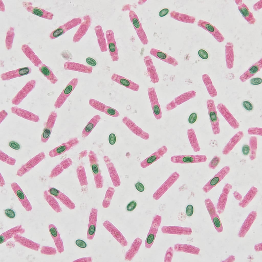
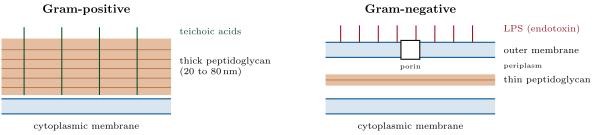
Every bacterium but a few is enclosed in a wall of peptidoglycan: chains of alternating N-acetylglucosamine and N-acetylmuramic acid, cross-linked by short peptides into a single molecule that surrounds the cell and resists the turgor pressure — several atmospheres — of the cytoplasm. Gram-positive bacteria have a thick wall of , many layers, threaded with teichoic acids; Gram-negative bacteria have a thin wall of one or two layers and, outside it, a second lipid bilayer, the outer membrane, whose outer leaflet is lipopolysaccharide (LPS, endotoxin — the molecule that the innate immune system of Chapter 15 recognises and that causes septic shock), enclosing a periplasm. Porins in the outer membrane admit small molecules and exclude many antibiotics. Outside may lie a polysaccharide capsule against phagocytes, flagella for swimming (Chapter 9) and pili for attachment and conjugation. Inside, the chromosome — one circular molecule of in most species — is folded into a nucleoid without a membrane, alongside plasmids. Some genera (Bacillus, Clostridium) can enclose a copy of the chromosome in a thick coat as an endospore, dehydrated and metabolically inert, which survives boiling, radiation and centuries of drought and germinates when nutrients return.
Method 12.2 (The Gram stain)
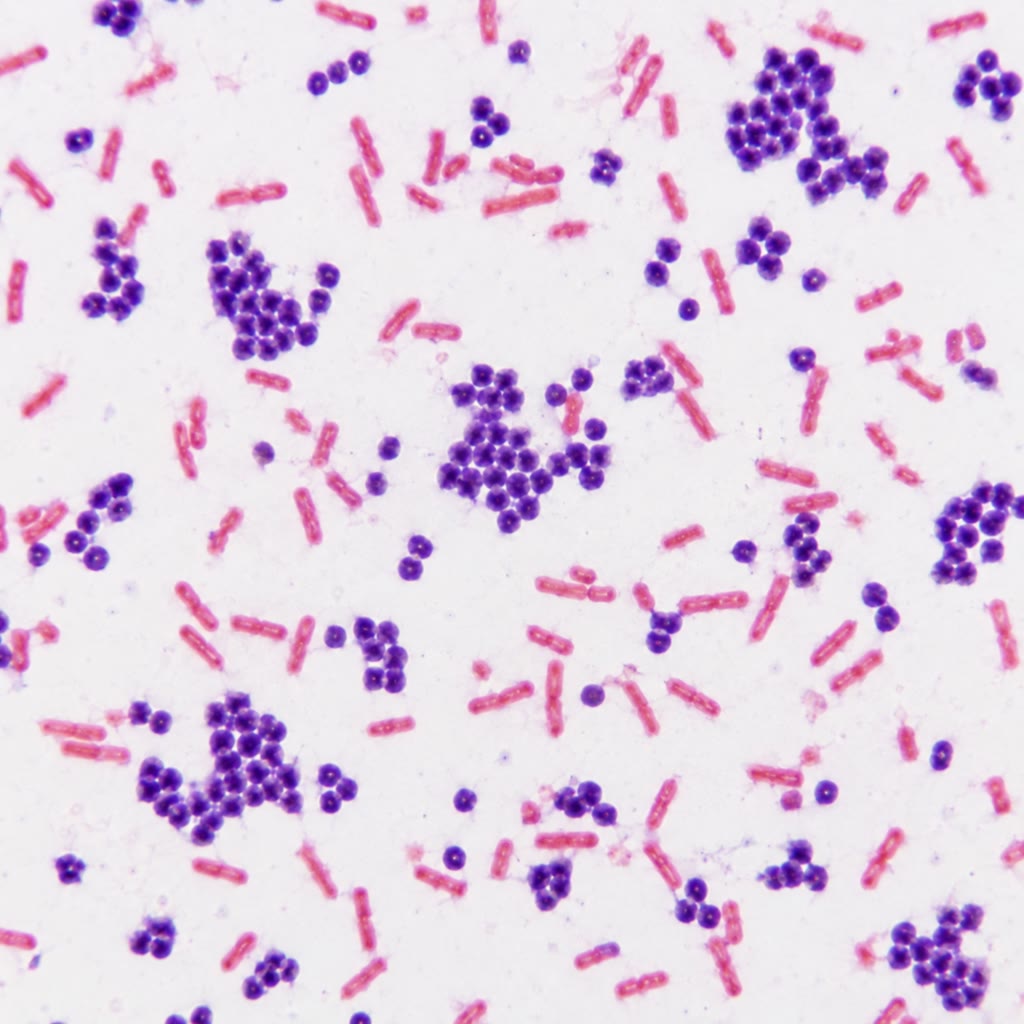
The oldest test in the bacteriological laboratory (Gram, 1884) still decides the first choice of antibiotic. (1) Smear and heat-fix the sample; (2) stain with crystal violet, then with iodine, which forms a large insoluble complex with the dye inside the cells; (3) wash with alcohol: the thick peptidoglycan of Gram-positives, dehydrated by the alcohol, traps the complex and the cells stay purple, while the thin wall of Gram-negatives lets it out; (4) counter-stain with safranin, which colours the now colourless Gram-negatives pink. Shape and arrangement — cocci in clusters (staphylococci) or chains (streptococci), rods, spirals — narrow the identification further, and a purple coccus in clusters from an abscess is a staphylococcus before any culture has grown.
12.2 Growth
Theorem 12.3 (Exponential growth, the Monod law and the chemostat)
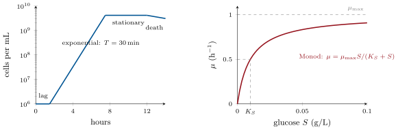
A population of cells dividing with generation time grows as with specific growth rate . The rate depends on the limiting nutrient by the Monod law
saturating at and half-maximal at . In a chemostat — a vessel of volume into which fresh medium of nutrient concentration flows at rate , and out of which culture leaves at the same rate, with dilution rate — the steady state has
where is the cell density and the yield (cells made per unit of nutrient). The experimenter sets the growth rate by turning a pump; the culture responds by adjusting the nutrient it leaves behind. If exceeds the cells cannot keep up and are washed out.
Proof. Cells: , growth minus outflow; at steady state with , , and inverting the Monod law gives . Nutrient: , inflow minus outflow minus consumption; at steady state, with , , whence . The steady state exists only if , that is ; beyond that the only steady state is , washout. Stability: if rises above it consumes more, falls below , falls below and declines — a negative feedback through the nutrient. ∎
Example 12.4 (A culture in numbers)
E. coli in rich medium at : , . From one cell, after twelve hours — cells, about , which is where a flask stops for want of oxygen and sugar; the “mass of the Earth in two days” is cells, and the arithmetic shows only that exponential growth never lasts. In a chemostat on glucose with , , and of cells per gram of glucose, a dilution rate of leaves of glucose and holds of cells, dividing every , indefinitely. The chemostat is how the physiology of a fixed growth rate is studied, and how thousands of generations of evolution are run in a bottle.
Definition 12.5 (Phases and states)
In a fresh flask (a batch culture) the cells first pause — the lag phase, while they induce the enzymes the new medium needs — then grow exponentially, then stop when a nutrient is exhausted or a waste accumulates: the stationary phase, in which the cells shrink, thicken their envelope, condense their nucleoid around protective proteins, switch on stress genes through the alternative sigma factor RpoS, and can survive for months; a death phase follows in which most cells lyse and a few live on the remains. Some species answer starvation with a developmental programme: Bacillus subtilis divides asymmetrically and the larger cell engulfs the smaller and builds the spore around it, a sequence of eight hours controlled by a cascade of sigma factors — the subunits that direct RNA polymerase to one set of promoters — alternating between the two compartments, the first bacterial example of differentiation and of cell-to-cell signalling through a wall.
12.3 Sensing one another
Definition 12.6 (Quorum sensing)
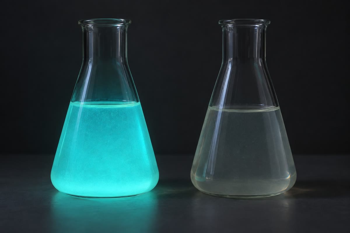
Bacteria detect their own density by quorum sensing: each cell secretes a small signal molecule, an autoinducer — acylated homoserine lactones in Gram-negatives, short peptides in Gram-positives — at a constant rate; the concentration in the medium rises with the number of cells, and when it crosses a threshold it binds a receptor that switches on a set of genes, among them the synthase of the autoinducer itself (hence the name). The genes so controlled are the ones that pay only in company: light in Vibrio fischeri, virulence factors in Staphylococcus aureus and Pseudomonas aeruginosa, biofilm matrix, competence for DNA uptake, and the killing of neighbours. Most bacterial sensing runs through two-component systems: a membrane histidine kinase that detects a stimulus and phosphorylates a cytoplasmic response regulator, which binds DNA — a cell has dozens, for phosphate, osmolarity, oxygen, and the autoinducers of other species.
Evidence. Nealson, Platt and Hastings (1970) grew the luminous marine bacterium Vibrio fischeri from a dilute inoculum and found that the cells made no light until they reached a density of some per millilitre, then all lit up together; medium in which a dense culture had grown, sterilised and added to a dilute culture, switched the light on at once. The cells were counting a secreted substance, not time or nutrient. The substance was identified as an acyl-homoserine lactone (Eberhard, 1981), and the genes — luxI for the synthase, luxR for the receptor, and the luciferase operon they control — were cloned into E. coli, which then glowed when crowded. In the squid that houses the bacterium at per millilitre, the light organ shines; in the sea, where the bacteria are dilute, they are dark and save the cost. ∎
Proposition 12.7 (A density threshold)
Let each of cells in a volume secrete autoinducer at rate molecules per second, and let the molecule be lost (degraded or diluted) with first-order rate . The concentration approaches , proportional to the cell density , with a time constant ; the receptor switches on when reaches its threshold , that is at the density
In an enclosed space — a squid’s light organ, an abscess, the mucus of a lung — is small because the signal is not carried away, so the threshold density is reached by fewer cells; in open water it is never reached. The cell therefore measures not its numbers but its numbers per unit of the volume it shares, which is what matters for any cooperative act, and the positive feedback of the autoinducer on its own synthase makes the switch sharp: once a few cells cross the threshold, their extra output pushes the rest across.
Proof. has the steady state and relaxes to it as . Setting and solving for gives the threshold density. ∎
12.4 The traffic of genes
Definition 12.8 (Horizontal gene transfer)
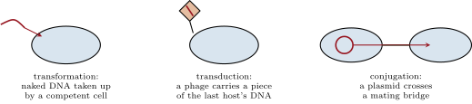
Bacteria reproduce clonally but exchange genes by three routes. Transformation: uptake of naked DNA from the environment by a competent cell — the route by which Griffith’s pneumococci acquired their capsule and Avery showed that the transforming principle was DNA (1944). Transduction: a phage that packages a piece of host DNA and injects it into the next host (Chapter 13). Conjugation: transfer of a plasmid through a pilus and a mating bridge from a donor carrying it to a recipient, demonstrated by Lederberg and Tatum (1946) with auxotrophic strains of E. coli that produced prototrophic recombinants only when mixed. Conjugative plasmids carry their own transfer genes; many also carry resistance genes, often clustered in integrons and flanked by transposons, so that a single mating can transfer resistance to five antibiotics at once, across species. The consequence is that a bacterial species has a core genome shared by all strains and an accessory genome that varies — a pangenome: two E. coli strains may differ by a fifth of their genes, and pathogenic strains differ from harmless ones mainly by acquired islands of virulence genes. The genes of the Year 2 volume’s mutation chapter arise by mutation; the genes of this one mostly arrive.
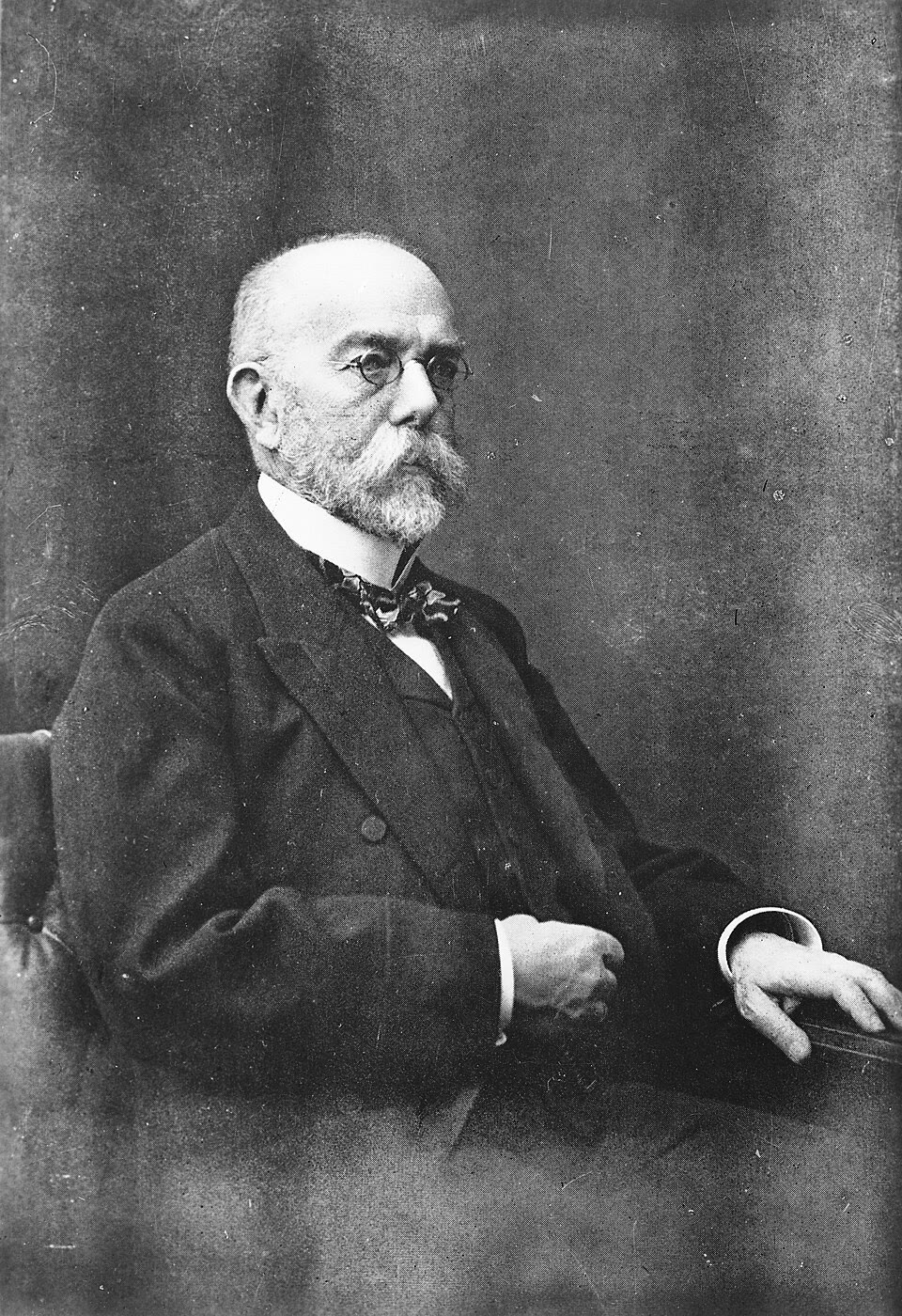
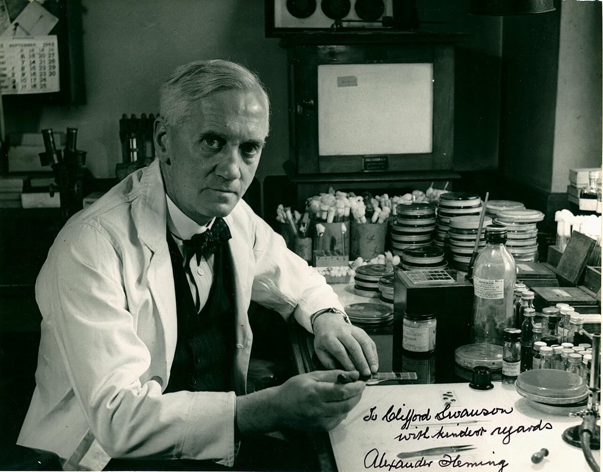
Evidence. Robert Koch (1876–1884) established that a specific bacterium causes a specific disease: he isolated Bacillus anthracis from sick animals, grew it in pure culture on solid medium (the agar plate and the single colony are his laboratory’s inventions), showed that the culture reproduced anthrax in healthy animals and could be re-isolated from them — the postulates that still define a pathogen — and went on to the tubercle bacillus and the cholera vibrio. Alexander Fleming (1928) noticed that a mould contaminating a plate of staphylococci had cleared a zone around itself; the substance, penicillin, was purified and shown to cure infections by Florey and Chain (1940–1941), and within a decade resistant staphylococci carrying a penicillin-destroying enzyme were common in hospitals — a warning Fleming himself gave in his Nobel lecture. ∎
12.5 Antibiotics and resistance
Definition 12.9 (Antibiotics)
An antibiotic is a molecule that kills bacteria or stops their growth at concentrations harmless to the host, by acting on a target the host lacks or has in a different form — selective toxicity. The targets are few. The cell wall: -lactams (penicillins, cephalosporins, carbapenems) mimic the terminal D-Ala-D-Ala of the peptidoglycan peptide and acylate the enzymes that cross-link it, so that the growing wall is weak and the cell bursts under its own turgor; vancomycin binds the D-Ala-D-Ala itself. The ribosome, whose bacterial form differs from the eukaryotic: aminoglycosides (the small subunit, causing misreading), tetracyclines (blocking tRNA entry), macrolides and chloramphenicol (the large subunit’s exit tunnel and peptidyl transferase). DNA gyrase and topoisomerase IV: the quinolones. RNA polymerase: rifampicin. Folate synthesis, which animals do not do: sulfonamides and trimethoprim. The membrane: the polymyxins, a last resort. The minimum inhibitory concentration (MIC) is the lowest concentration of a drug that prevents visible growth of a strain; a strain is called resistant when its MIC exceeds the concentration the drug reaches in the patient.
Method 12.10 (Measuring susceptibility)
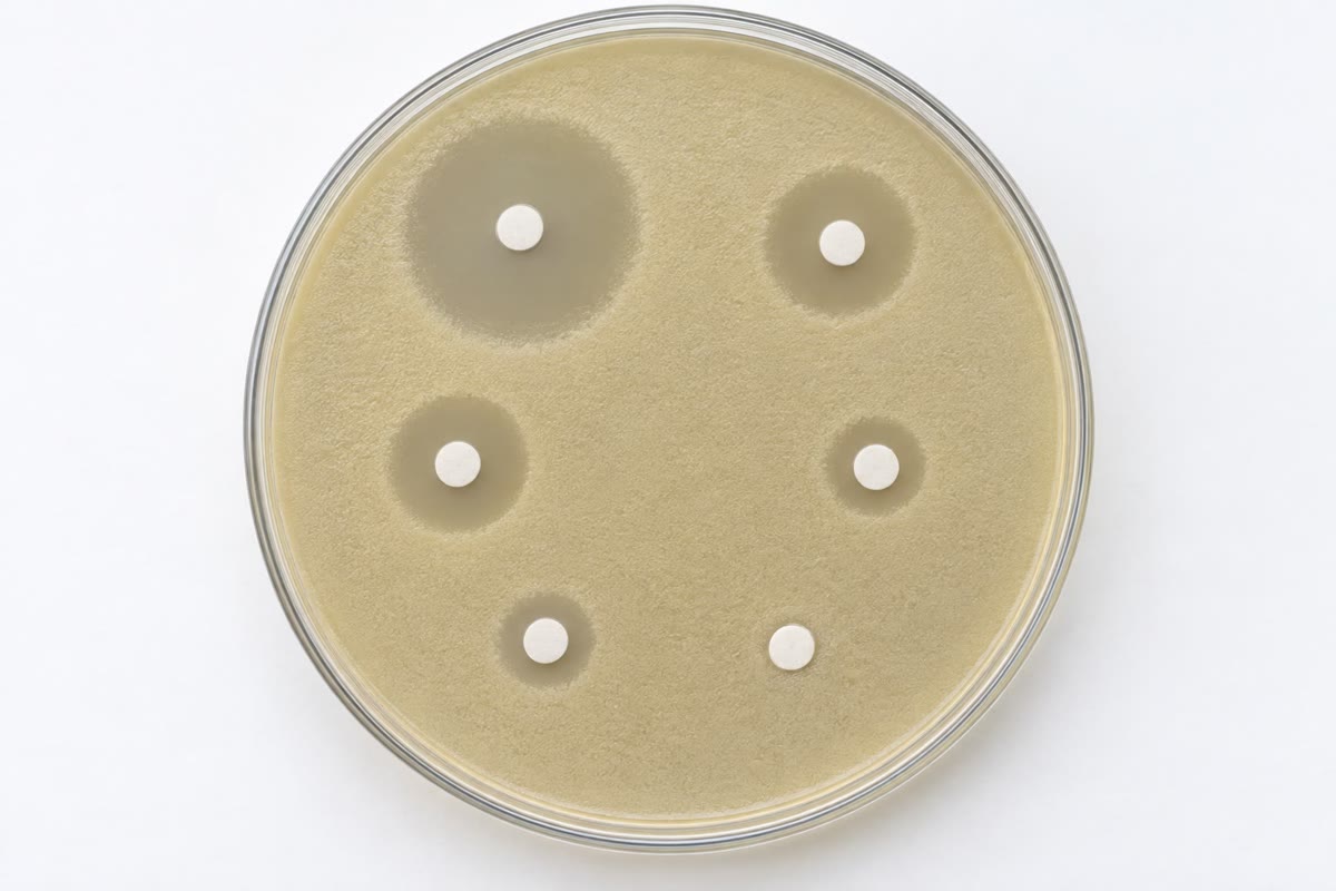
Two tests. Broth dilution: inoculate a fixed number of cells (about per millilitre) into a row of tubes or wells containing the antibiotic in twofold steps, incubate overnight, and read the first clear well: that concentration is the MIC. Disk diffusion: spread the strain on an agar plate, place paper discs loaded with known amounts of several antibiotics, incubate; the drug diffuses outward, its concentration falling with distance, and growth is inhibited out to the radius where the concentration equals the MIC, so that the diameter of the clear zone — read against calibrated tables — gives the susceptibility for each disc on one plate. Both tests take a day; sequencing the genome for known resistance genes and mutations takes the same day and is beginning to replace them.
Definition 12.11 (Resistance)
A bacterium resists an antibiotic by one of five means. It destroys the drug: -lactamases hydrolyse the -lactam ring, and the extended-spectrum and carbapenemase variants now destroy the newest ones. It alters the target: a mutation in the ribosomal protein or gyrase that the drug binds, or, in MRSA (methicillin-resistant Staphylococcus aureus), an acquired gene for a cross-linking enzyme that -lactams do not bind; vancomycin resistance replaces the terminal D-Ala with D-lactate, which the drug does not recognise. It pumps the drug out with efflux pumps, often broad in specificity. It stops the drug getting in, by losing a porin. Or it bypasses the blocked step with an alternative enzyme. Target mutations arise by chance in the patient at rates of to per division and are selected during treatment; the enzymes and pumps are mostly acquired on plasmids from the vast reservoir of soil and gut bacteria, in which they evolved over ages against the antibiotics that fungi and other bacteria make. Persisters — dormant cells that survive a drug without being genetically resistant — explain relapses after treatment that should have worked.
Proposition 12.12 (Why resistance is inevitable and combination is the answer)
An infection of cells treated with a drug against which resistance mutations arise at per division already contains about ten resistant cells; the drug kills the rest and the ten take over — the arithmetic of Chapter 11 exactly, since a bacterial population and a tumour are both evolving clones under selection. Two drugs with independent targets face a doubly resistant cell at per division, which a billion cells do not contain. This is why tuberculosis, whose lesions hold – bacilli, has been treated with three or four drugs at once since the 1950s and why monotherapy of it produced resistance within months; it is also why every use of an antibiotic, on a patient or in a feedlot, selects resistance somewhere, and why the rate of new antibiotics — none of a new class against Gram-negatives since the 1960s — is the measure against which the losses are counted. The remedies are those of any evolutionary contest: use less, combine, finish the course so that partially resistant cells do not survive to breed, and find new targets.
Example 12.13 (The mutant selection window)
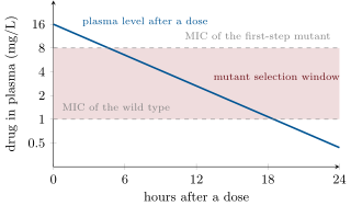
A strain has MIC ; its first-step resistant mutants, present at one in , have MIC . A dose that holds the drug at kills the wild type and leaves the mutants to grow without competition: the concentration range between the MIC of the wild type and that of the mutants is the mutant selection window, and a treatment that sits in it breeds resistance. A dose above kills both; too low a dose, or a course stopped when the patient feels better and the drug level is falling through the window, is how a resistant population is made. The same logic sets the regimens of tuberculosis treatment: six months, four drugs, at doses above every single-step mutant’s MIC.
Remark 12.14 (Most bacteria are not pathogens)
The bacteria of this chapter’s clinic are a tiny minority. Of the estimated million species, a few hundred cause human disease; the rest run the cycles of nitrogen, sulfur and carbon (the Year 2 volume), fix the nitrogen of every legume, digest the cellulose in every ruminant, and make up the microbiomes of Chapter 14, whose loss under antibiotic treatment is a cost of every course. The antibiotics themselves are their molecules — weapons and signals evolved among soil bacteria and fungi long before there were hospitals — and so are most of the resistance genes.
12.6 Exercises
Exercise 12.1 ★
Describe the Gram-positive and Gram-negative envelopes and explain why the Gram stain distinguishes them.
Solution
Solution of Exercise 12.1.
Gram-positive: cytoplasmic membrane wrapped in a thick, many-layered peptidoglycan wall threaded with teichoic acids. Gram-negative: a thin peptidoglycan layer in a periplasm, then an outer membrane whose outer leaflet is lipopolysaccharide, with porins. The crystal violet–iodine complex is trapped in the thick, alcohol-dehydrated wall of the Gram-positive (purple) but washed out of the Gram-negative through its thin wall and disrupted outer membrane, which then takes the pink counter-stain.
Exercise 12.2 ★
A culture of cells per millilitre reaches in of exponential growth. Find the number of generations, the generation time and .
Solution
Solution of Exercise 12.2.
-fold : about generations; ; .
Exercise 12.3 ★
Name the three routes of horizontal gene transfer and give the classic experiment that demonstrated each.
Solution
Solution of Exercise 12.3.
Transformation — Griffith (1928) and Avery, MacLeod and McCarty (1944): heat-killed virulent pneumococci transform live avirulent ones, and the agent is DNA. Transduction — Zinder and Lederberg (1952): phage P22 carries Salmonella genes from one strain to another through a filter that stops cells. Conjugation — Lederberg and Tatum (1946): two auxotrophic E. coli strains give prototrophic recombinants only when mixed, requiring cell contact (Davis’s U-tube).
Exercise 12.4 ★
Give the target of penicillin, of streptomycin, of ciprofloxacin and of rifampicin, and say why each is selectively toxic.
Solution
Solution of Exercise 12.4.
Penicillin: the transpeptidases that cross-link peptidoglycan, which animal cells do not have. Streptomycin: the small ribosomal subunit, whose bacterial form differs from the eukaryotic. Ciprofloxacin: DNA gyrase, a bacterial topoisomerase absent from humans. Rifampicin: the subunit of bacterial RNA polymerase, which the eukaryotic enzyme does not share.
Exercise 12.5 ★★
A chemostat has , , , . Compute and at , and h, and the dilution rate at which washout begins.
Solution
Solution of Exercise 12.5.
, . : , . : , . : , . Washout when .
Exercise 12.6 ★★
Using Proposition 12.7, compare the threshold density for quorum sensing in a squid light organ () and in flowing sea water (), with molecules per cell per hour and ( molecules per litre).
Solution
Solution of Exercise 12.6.
. Light organ: per litre per mL. Sea water: per litre per mL — a thousand times higher, never reached in the sea.
Exercise 12.7 ★★
A disc-diffusion plate shows zones of , and for three drugs. Interpret each, and explain why a zone diameter can be converted into an MIC.
Solution
Solution of Exercise 12.7.
: sensitive, low MIC. : reduced susceptibility, intermediate — may fail at ordinary doses. : resistant, growth up to the disc. The drug diffuses from the disc so that its concentration falls with distance in a reproducible way; growth stops where the concentration equals the MIC, so zone radius and MIC are related by a calibration curve established with strains of known MIC.
Exercise 12.8 ★★
Explain how a single conjugation event can make a sensitive Klebsiella resistant to five antibiotics, and why resistance genes are so often found together on plasmids.
Solution
Solution of Exercise 12.8.
The conjugative plasmid carries an integron whose cassette array holds several resistance genes, plus transposons carrying more; the plasmid crosses in one mating and expresses them all. They cluster because selection for any one of the drugs maintains the whole plasmid, so genes that hitch-hike on it persist (co-selection); integrons capture cassettes; transposons hop onto plasmids; and the plasmid, not the gene, is the unit of transfer.
Exercise 12.9 ★★
A patient has bacteria and takes a drug against which resistance arises at per division. Compute the expected resistant cells at the start; then with two independent drugs. Why are persisters a different problem?
Solution
Solution of Exercise 12.9.
Drug alone: resistant cells expected. Two independent drugs: . Persisters are not mutants: a small fraction of dormant cells survives any drug and regrows into a sensitive population when the drug is gone, so a second drug does not help; only a treatment long enough to catch them as they wake, or a drug active on dormant cells, does.
Exercise 12.10 ★★★
Show that the chemostat steady state is stable: perturb slightly above and follow the signs of and . Then explain why two species competing for one nutrient in a chemostat cannot coexist at steady state, and which one wins at a given .
Solution
Solution of Exercise 12.10.
: consumption exceeds supply, so falls below ; then and , falls back; as falls, rises again. Both deviations are corrected: the steady state is stable. Two species: each needs to persist, which fixes its own ; the nutrient can take only one value, so at most one species persists. The one with the lower drives below what the other needs, and the other is washed out; which species that is can change with if their Monod curves cross.
Exercise 12.11 ★★★
The autoinducer induces its own synthase. Modify the model of Proposition 12.7 so that jumps from to when , and show that the system can have two stable states over a range of densities (hysteresis): a population that has switched on stays on at a density where a naive population would be off. What is the biological use of that?
Solution
Solution of Exercise 12.11.
With density , the off state exists while it stays below , i.e. ; the on state exists while it stays above, i.e. . For both are self-consistent: a population that has switched on keeps itself on with its higher output, one that has not stays off. Use: commitment — once a colony has decided to build a biofilm or release toxin, a dip in density does not undo the decision, and the switch is sharp and synchronous rather than graded.
Exercise 12.12 ★★★
Explain the mutant selection window and use it to argue for or against each of: (a) high doses for short courses; (b) low doses for long courses; (c) stopping when symptoms resolve; (d) combination therapy.
Solution
Solution of Exercise 12.12.
(a) High doses for short courses: the level stays above the mutant MIC and kills wild type and first-step mutants alike — favoured, limited by toxicity. (b) Low doses for long courses: the level sits in the window for days and breeds resistance — against. (c) Stopping when symptoms resolve: the survivors are the least sensitive cells and the falling level passes through the window — against. (d) Combination: a cell must be resistant to both, which a billion cells do not contain — for.
12.7 Problem: A Chemostat and a Clinic
Problem 12.1
Weekend problem — a culture grown in a flask and held in a chemostat, a luminous bacterium counted to its quorum, and an infection treated against the arithmetic of resistance, ending on the cells in the chemostat, the density at which the light comes on and the odds that a two-drug course meets a resistant cell
Data: E. coli at , ; a cell weighs . Chemostat: , , , , glucose, . Vibrio: autoinducer molecules per cell per hour, molecules per litre (), in the light organ. Infection: cells; resistance to drug A at per division, to drug B at ; wild-type MIC of A , first-step mutant ; a dose gives falling with half-life .
Part I — The flask.
- One cell in of broth. How many cells after of exponential growth, and what density per mL?
- The broth supports at most cells per mL. When does growth stop, and what is the culture’s mass of cells?
- How long would it take the descendants of one cell to reach the mass of the Earth ( g)? Comment.
- The lag phase lasts when cells from stationary phase are inoculated into fresh medium, and almost nothing when exponential cells are used. Explain.
- Compute from . If the medium is switched to a sugar on which , what is , and by what factor does the number of cells after fall?
- A sample of the stationary culture is spread on a plate and gives colonies from of a dilution. What is the viable count, and why might it differ from a microscope count?
Part II — The chemostat.
- Compute the dilution rate and the generation time the culture is forced to adopt.
- Compute and (in g/L and cells per mL).
- How many cells leave the vessel per hour? How many generations does the culture go through in a month?
- The pump is turned up to . New and ? What happens at ?
- A mutant appears with and the same . At , what does it need, and what happens to the original strain? (Use Exercise 12.10.)
- A month’s run is generations. If a beneficial mutation arises at per division and the vessel holds cells, how many beneficial mutations arise per generation? What does this say about evolution in a chemostat?
Part III — The quorum.
- Compute the threshold cell density for light in the light organ, in cells per mL.
- The light organ holds of bacteria at per mL. By what factor is the autoinducer above threshold?
- In sea water the signal is swept away with . Threshold density? Compare with the cells per mL of the open sea.
- After the squid expels of the bacteria at dawn, how long until the remaining per mL, dividing every , restore per mL? Does the light go out meanwhile?
- Each glowing cell spends about of its energy on light. Why is a dark cell in the sea favoured, and why not in the organ?
- A pathogen uses the same system to delay its toxins until it is numerous. Propose a drug strategy that exploits this and say why it might select resistance more slowly than an antibiotic.
Part IV — The clinic.
- Expected number of cells resistant to A at the start of treatment; to B; to both.
- Probability that no cell resistant to A exists; to both.
- After the dose, when does the drug level fall to , and to ? How long per dose does it spend in the mutant selection window?
- If the drug is given every , what fraction of the time is the level in the window, and what does that predict over a ten-day course with A alone?
- Suggest a dosing change that keeps the level above throughout (compute the interval or the dose needed), and state its cost.
- One cell in is a persister, dormant and untouched by any concentration of either drug. How many survive the course, what happens when the drug is withdrawn, and what does this imply for the length of treatment?
- Summarise: the cells per mL in the chemostat (question 8), the light threshold density (question 13), and the probability that the two-drug course meets no resistant cell (question 20).
Solution
Solution of Problem 12.1.
1. generations: cells, per mL. 2. cells : after ; mass . 3. cells: generations, . Nutrients, oxygen and waste stop it within a day; exponential growth is always brief. 4. Stationary cells have dismantled ribosomes and repressed their metabolic enzymes; they must rebuild them before dividing. Exponential cells arrive fully equipped. 5. ; at , ; after instead of : a factor fewer. 6. viable cells per mL. The microscope also counts dead cells and cells that will not grow on the plate, and a clump gives one colony. 7. ; . 8. ; cells per litre, per mL. 9. cells per hour; generations a month. 10. : , . At , : washout. 11. The mutant holds ; at that glucose the original grows at and declines as : washed out over a few weeks. The mutant takes over. 12. beneficial mutations per generation: adaptation is continuous, with a new advantageous clone sweeping every few hundred generations — the chemostat is an evolution machine. 13. per litre cells per mL. 14. -fold. 15. per litre, per mL — six orders of magnitude above the per mL of the sea: dark. 16. Tenfold is doublings, . At per mL the population is still times above threshold: the light stays on. 17. In the sea no one sees the light and it feeds no host: a dark mutant saves and outgrows the glowers. In the organ the squid selects for light — it expels or fails to feed dark cheaters — so the cost buys a home. 18. Block the receptor or destroy the autoinducer (quorum quenching): the bacteria live but never deploy their toxins, and the immune system clears them. Since sensitive and “resistant” bacteria grow equally — nothing is killed — the selective advantage of escaping the drug is small, and resistance should spread more slowly than to a killing drug. 19. A: ; B: ; both: . 20. ; . 21. : one half-life, ; : four, ; in the window from to , per dose. 22. Dosing every : in the window from hour to hour , two thirds of the time; over ten days the first-step mutants of A — ten of them at the start — are selected and A alone fails. 23. Above throughout: dose every half-life (), or a dose peaking at every ( in three half-lives), or a continuous infusion. Costs: toxicity of high peaks, or six doses a day that patients will not keep. 24. persisters survive whatever the drugs; when treatment stops they wake and regrow a fully sensitive population — a relapse. Treatment must last long enough to catch them as they leave dormancy, which is why tuberculosis takes six months. 25. cells per mL in the chemostat; the light comes on at cells per mL; that the two-drug course meets no resistant cell.