University Biology — Year 1 · Bachelor Year 1
10Carbohydrates
A potato and a sheet of paper are made of the same molecule, glucose, strung into chains. One is a meal and the other is not, because the chains are joined by two different bonds: the link of starch coils the chain into a helix that our enzymes open, the link of cellulose stretches it into ribbons that stack into fibres no mammal can digest. A liver stores a hundred grams of glucose as glycogen, a tree holds itself up with cellulose, a beetle wears chitin, and every cell displays sugars on its surface as its identity. This chapter describes the simple sugars, the bonds that join them, the polysaccharides they build, and the sugar-decorated molecules of the cell surface and the plant wall.
10.1 Monosaccharides
Definition 10.1 (Monosaccharide)
Carbohydrates are molecules of composition and their derivatives. A monosaccharide (simple sugar) is a chain of three to seven carbons, one of which carries a carbonyl group and the others hydroxyls: an aldose when the carbonyl is terminal (an aldehyde: glucose, galactose, ribose), a ketose when it is internal (a ketone: fructose). By length: trioses (: glyceraldehyde), pentoses (: ribose, deoxyribose), hexoses (: glucose, fructose, galactose). Every carbon bearing four different groups is a chiral centre, so each formula stands for several stereoisomers: glucose has four chiral carbons and sixteen isomers, of which living things use one, D-glucose. Epimers differ at one carbon only (glucose and galactose at C4).
Proposition 10.2 (Ring forms and anomers)
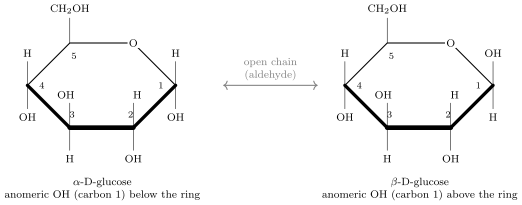
In water a pentose or hexose is almost entirely in a ring: the carbonyl carbon reacts with a hydroxyl of the same molecule (C5 in glucose) to close a six-membered pyranose ring or a five-membered furanose ring (fructose, ribose). The carbonyl carbon becomes a new chiral centre, the anomeric carbon, whose hydroxyl can lie below the ring plane ( anomer) or above it ( anomer) in the Haworth drawing; the two forms interconvert through the open chain in minutes. In solution, glucose is , and less than open chain. The free anomeric carbon is what makes a sugar reducing: it can open and reduce a copper or silver reagent.
Method 10.3 (Reading a Haworth projection)
- Find the ring oxygen; the anomeric carbon (C1 in an aldose, C2 in a ketose) is the ring carbon to its right, bearing both a hydroxyl and the ring oxygen.
- The carbon carrying the group outside the ring (C5 in glucose) is to the left of the ring oxygen; its points up in a D-sugar.
- Anomeric hydroxyl down: ; up (on the same side as ): .
- Compare the other hydroxyls with those of glucose (down, up, down for C2, C3, C4): a single difference identifies an epimer (galactose: C4 up).
Example 10.4 (Sugars that are not )
Deoxyribose lacks the hydroxyl at C2 (Chapter 11); glucosamine and N-acetylglucosamine carry an amine instead of the C2 hydroxyl (chitin, peptidoglycan); glucuronic acid has a carboxyl at C6 (the matrix polysaccharides); sugar phosphates (glucose-6-phosphate, ribose-5-phosphate) are the forms in which sugars enter metabolism; sugar alcohols (glycerol, sorbitol) have no carbonyl. The core is the same: a small polyhydroxylated carbon chain that water loves.
10.2 The glycosidic bond
Definition 10.5 (Glycosidic bond, disaccharide)
A glycosidic bond joins the anomeric hydroxyl of one sugar to a hydroxyl of another (or of an alcohol, an amine, a base) by condensation, fixing the anomeric carbon in its or configuration; it is named by the configuration and the carbons joined: , , . Two sugars so joined form a disaccharide: maltose (glucose glucose, from starch digestion), lactose (galactose glucose, the sugar of milk), sucrose (glucose fructose, the sugar of plant sap and of the kitchen). In sucrose both anomeric carbons are engaged: it is non-reducing and does not open, which is why plants transport it (Chapter 24).
Method 10.6 (Testing for a reducing sugar)
- Heat the solution with an alkaline copper(II) reagent (Benedict’s or Fehling’s).
- A free anomeric carbon opens to an aldehyde, reduces the blue to a brick-red precipitate of ; the depth of colour scales with the amount of sugar.
- Glucose, fructose, maltose, lactose: positive. Sucrose: negative — unless first hydrolysed by acid or by the enzyme invertase, after which its glucose and fructose react.
- Starch does not react (its single reducing end is one in thousands); it is detected with iodine, which enters the helix of amylose and turns blue-black.
10.3 Polysaccharides
Definition 10.7 (Polysaccharide)
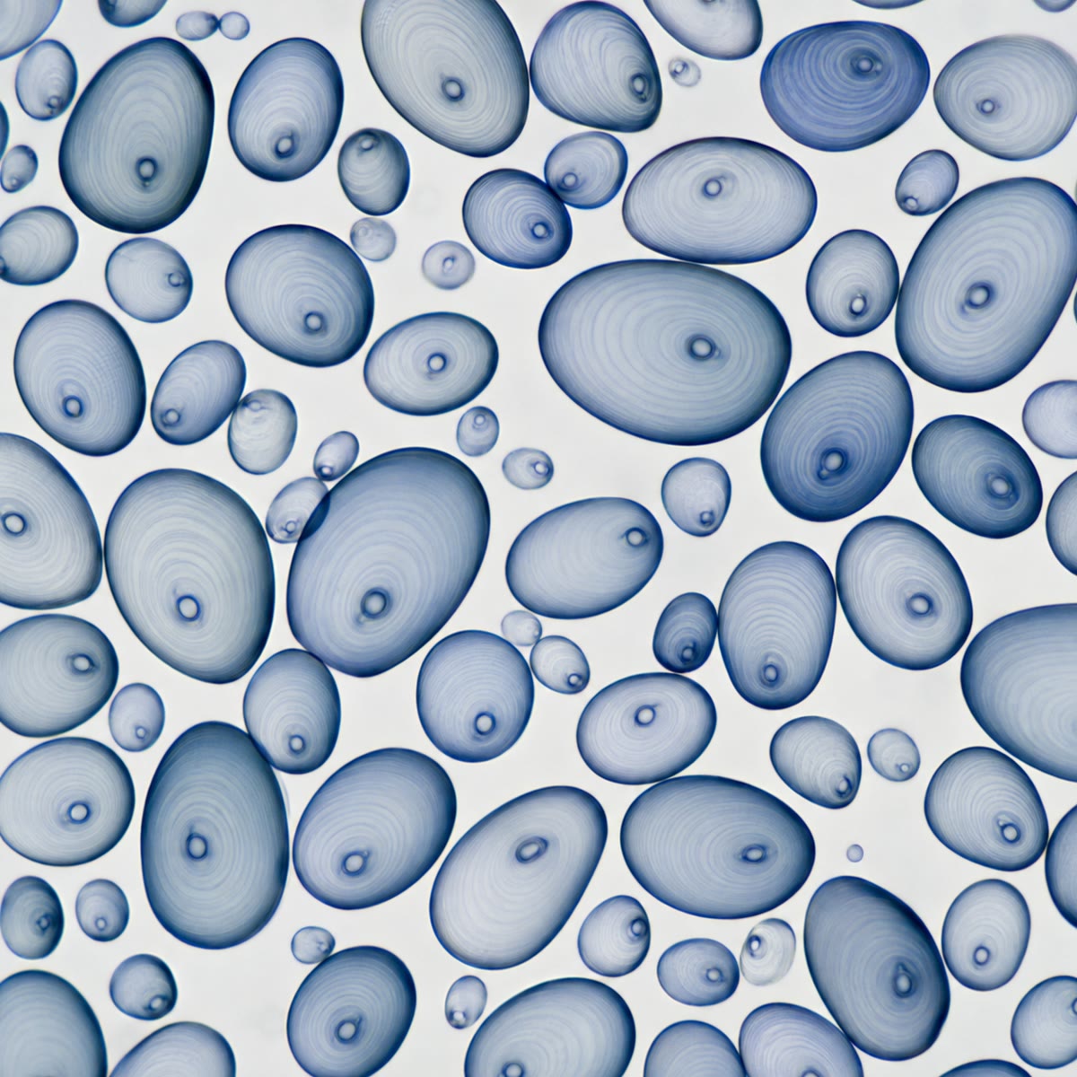
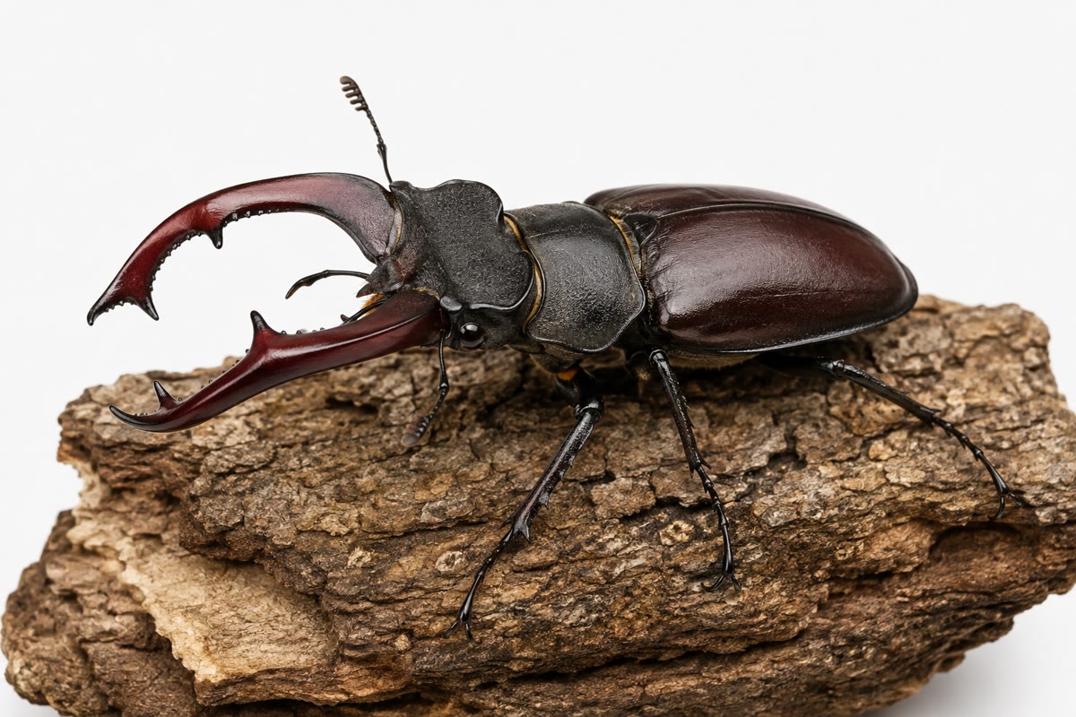
A polysaccharide is a chain of hundreds to tens of thousands of monosaccharides joined by glycosidic bonds, linear or branched. Storage polysaccharides: starch in plants — amylose, unbranched glucose, , and amylopectin, with branches every residues, — packed in grains; glycogen in animals and fungi, like amylopectin but branched every residues, in cytosolic granules of the liver and muscles. Structural polysaccharides: cellulose, unbranched glucose, residues, in plant walls; chitin, N-acetylglucosamine, in the walls of fungi and the cuticles of arthropods; peptidoglycan, alternating N-acetylglucosamine and N-acetylmuramic acid cross-linked by short peptides, the one-molecule mesh that is the bacterial wall.
Proposition 10.8 (The bond decides the shape, and the shape the function)
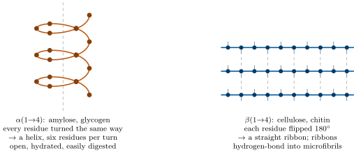
In an chain every glucose is turned the same way as the last and the chain curls into a helix of six residues per turn, an open, hydrated coil that enzymes enter easily: the form of a store. In a chain each glucose is flipped relative to its neighbour and the chain is a straight, flat ribbon; ribbons lie side by side and hydrogen-bond into microfibrils of thirty to a hundred chains, crystalline, insoluble, with the tensile strength of steel per unit mass: the form of a fibre. The enzymes that hydrolyse bonds (amylases) cannot touch bonds; cellulases exist in bacteria, fungi and a few animals, and the mammals that live on grass do so through symbionts (Chapter 22).
Example 10.9 (Why glycogen is branched)
Glycogen is degraded from its non-reducing ends, one glucose at a time, by glycogen phosphorylase. An unbranched chain of glucoses would have one such end and release one glucose per enzyme turnover; a glycogen particle of the same size, branched every twelve residues, has some ends and releases two thousand at once. Branching also keeps the particle compact and soluble. The liver’s hundred grams of glycogen can be mobilised at ten grams an hour; the same glucose stored as one long chain would take years to unwind.
Example 10.10 (Why not store glucose itself)
A hundred grams of glucose () dissolved in the of water of a liver’s cells would add to a cytosol at : the cells would swell to three times their volume and burst. As glycogen the same glucose is particles, , osmotically invisible. A polymer is a way of storing a great many molecules as one.
10.4 Glycoconjugates and the plant wall
Definition 10.11 (Glycoproteins, proteoglycans, glycolipids)
Sugars are attached covalently to proteins and lipids to form glycoconjugates. A glycoprotein carries short branched chains (a dozen sugars) on some of its asparagine, serine or threonine residues, added in the ER and Golgi (Chapter 6): nearly every secreted and membrane protein is one. A proteoglycan is a protein core bearing long unbranched chains of repeating acidic disaccharides, the glycosaminoglycans (hyaluronan, chondroitin sulfate, heparin), which bind enormous amounts of water and give cartilage, the vitreous body and the extracellular matrix their resilience. Glycolipids carry sugars on a lipid tail in the outer leaflet of the plasma membrane. Together with the sugars of glycoproteins they form the glycocalyx, the sugar coat by which cells are recognised.
Example 10.12 (Blood groups)
The ABO blood groups are three versions of one sugar chain on the red cell’s surface glycolipids and glycoproteins: the O chain ends in fucose; A adds an N-acetylgalactosamine to it, B adds a galactose, by two versions of one enzyme; AB cells carry both. A single sugar residue is enough for the immune system to distinguish self from foreign, and for a transfusion to succeed or kill.
Proposition 10.13 (The plant cell wall)
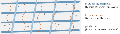
The primary wall of a plant cell is a fibre-reinforced composite: cellulose microfibrils (a quarter of the mass, tensile strength) laid in layers, tied together by hemicelluloses (branched polysaccharides that hydrogen-bond to the microfibrils and bridge them) and embedded in a gel of pectins (acidic polysaccharides that hold water and calcium and cement cells together in the middle lamella), with a few percent of protein. It is thin (), permeable to water and small solutes, and strong enough to hold of turgor. Cells that stop growing may add a thick secondary wall with more cellulose and lignin, an aromatic polymer that waterproofs and stiffens it: wood is secondary walls.
10.5 Sugars as fuel and carbon
Proposition 10.14 (The place of glucose)
Carbohydrates yield on oxidation and are the fuel every cell can use, with or without oxygen (Chapter 15); glucose is the sugar of the blood (), sucrose that of plant sap, and starch and glycogen their stores. Sugars are also the carbon from which the cell makes everything else: the pentoses of the nucleotides, the glycerol of lipids, the carbon skeletons of amino acids all leave the sugar pathways (Chapter 16). Photosynthesis makes sugar first; the rest follows.
Example 10.15 (A day’s sugar)
A human eats some of carbohydrate a day, nearly all as starch and sucrose; the gut hydrolyses it to glucose, fructose and galactose, the liver turns the last two into glucose, and glucose circulates at in the whole blood — twenty minutes’ supply for the brain, which takes a day, refilled continuously from the liver’s glycogen. The lens of the eye, the red cells and the kidney medulla live on glucose alone; nothing else in the diet can replace it, and when it runs out the liver makes it from protein.
10.6 Exercises
Exercise 10.1 ★
Classify glucose, fructose, ribose and glyceraldehyde by number of carbons and by aldose or ketose.
Exercise 10.2 ★
What is an anomeric carbon, and why does a disaccharide with both anomeric carbons in the bond not react with Benedict’s reagent?
Solution
Solution of Exercise 10.2.
The carbonyl carbon after ring closure, bearing the ring oxygen and a hydroxyl, which can open back to the aldehyde or ketone. In sucrose both anomeric carbons are locked in the glycosidic bond; neither can open, so no aldehyde forms and the copper is not reduced.
Exercise 10.3 ★
Name the monomers and bonds of starch, glycogen, cellulose and chitin.
Exercise 10.4 ★
From the Haworth figure, list the positions (up or down) of the hydroxyls on carbons 1 to 4 of -D-glucose, then draw or describe -D-galactose.
Solution
Solution of Exercise 10.4.
C1 down, C2 down, C3 up, C4 down. -D-galactose is the same with the C4 hydroxyl up.
Exercise 10.5 ★★
Explain, from the geometry of the and bonds, why amylose forms a helix and cellulose a ribbon, and why the iodine test works on the first and not the second.
Solution
Solution of Exercise 10.5.
In the bond the anomeric hydroxyl points down, and each residue can be added at the same angle as the last: the chain turns steadily and closes into a helix. In the bond it points up, and a residue can only be added flipped over: the chain runs straight. Iodine molecules fit inside the amylose helix and their electronic state changes (blue); a flat ribbon offers no cavity.
Exercise 10.6 ★★
Three tubes hold glucose, sucrose and starch. Design a sequence of tests (Benedict, iodine, acid hydrolysis) that identifies each, giving the expected result of every test.
Solution
Solution of Exercise 10.6.
Iodine: only starch turns blue-black. Benedict on the other two: only glucose gives a red precipitate; sucrose stays blue. Confirm sucrose by heating with dilute acid, neutralising, and repeating Benedict: now positive.
Exercise 10.7 ★★
A glycogen particle holds glucose residues branched every 12. Estimate the number of non-reducing ends and compare with an amylose molecule of the same size. If phosphorylase releases one glucose per second from each end, how long does each take to release of its glucose?
Solution
Solution of Exercise 10.7.
Chains of 12 residues: about chains, half of them outermost: some ends. Amylose: one end. To release glucoses at one per second per end: glycogen ; amylose , nearly an hour and a half.
Exercise 10.8 ★★
Cellulose microfibrils have a tensile strength of about . A cell of diameter and wall thick holds a turgor of . Compute the wall stress ( for a sphere) and compare with the strength.
Solution
Solution of Exercise 10.8.
: a fiftieth of the fibril strength; the wall has a large safety margin, needed because the fibrils are only a quarter of the wall.
Exercise 10.9 ★★
Lactose intolerance: adults who lack intestinal lactase get cramps and diarrhoea after milk. Explain, using the bond that lactase breaks, what happens to the undigested lactose in the colon (bacteria, osmosis).
Solution
Solution of Exercise 10.9.
Lactase breaks the bond between galactose and glucose; without it lactose is not absorbed (only monosaccharides cross the epithelium). In the colon bacteria ferment it to acids and gas (cramps, bloating), and the undigested sugar and acids draw water osmotically into the lumen: diarrhoea.
Exercise 10.10 ★★★
A cow eats of grass dry matter a day, of it cellulose. It has no cellulase. Explain how it nevertheless obtains energy from the cellulose, why the process must occur before the small intestine, and estimate the energy at stake if of the cellulose is fermented and yields of usable products per gram.
Solution
Solution of Exercise 10.10.
Bacteria and protists in the rumen secrete cellulases and ferment the glucose to short fatty acids, which the cow absorbs and oxidises; the microbes themselves are later digested. The fermentation must precede the small intestine so that its products reach the absorbing surface and the microbial protein reaches the stomach and intestine. Cellulose: ; fermented ; a day, most of the cow’s energy.
Exercise 10.11 ★★★
The A and B blood-group antigens differ by one sugar added by two versions of a transferase; type O has neither. Explain why an O person can donate red cells to anyone but receive only from O, and what an A person’s plasma must contain.
Solution
Solution of Exercise 10.11.
O cells carry neither antigen and provoke no antibody in anyone; O plasma, however, holds antibodies against both A and B, so an O person attacks any A or B cell received. An A person’s immune system has never seen B and makes anti-B antibodies (against gut bacteria bearing similar sugars), but tolerates A: A plasma contains anti-B only.
Exercise 10.12 ★★★
“Glucose is the universal fuel, cellulose the universal building material, and they are the same molecule.” Discuss in a paragraph: what the sentence gets right, what one bond changes, and what it says about the relation between chemistry and biology.
Solution
Solution of Exercise 10.12.
Right: glucose is the fuel of nearly every cell and cellulose the most abundant organic molecule on Earth, and both are polymers of, or the same as, D-glucose. One bond changes everything: gives a coil that enzymes open and cells burn; gives a fibre that resists enzymes, water and tension, and that most animals cannot use at all. Biology is not the list of molecules but the geometry of their joining — the same chemistry can be food or scaffold according to a single stereochemical choice, and organisms are separated by which enzymes they possess to undo it.
10.7 Problem: The Tree of Glycogen
Problem 10.1
Weekend problem — a glycogen particle counted branch by branch, the liver’s store weighed and its release timed, the osmotic price that polymerisation avoids, ending on the glucose release rate of the liver
A glycogen particle is a tree of chains: an inner chain of glucose residues bears two branches, each of residues and each bearing two branches in turn, and so on for tiers; the outermost tier’s chains are unbranched. Each glucose residue in the polymer has a mass of . A resting human liver of holds of glycogen in of cell water, and releases glucose into the blood at between meals. Glycogen phosphorylase removes one glucose from a non-reducing end per turnover, at turnovers per second per enzyme molecule.
Part I — Counting the tree.
- How many chains does tier contain ( for the inner chain)? Give the number for tiers 1, 2, 3 and 12.
- Compute the total number of chains in the particle.
- Compute the total number of glucose residues.
- Compute the mass of the particle in daltons and in grams.
- What fraction of all the chains are in the outermost tier? How many non-reducing ends does the particle offer?
- An amylose molecule of the same number of residues has how many non-reducing ends?
- If each tier adds to the radius, compute the particle’s diameter and compare it with a ribosome ().
Part II — The liver’s store.
- Compute the number of particles in the liver.
- Compute the glucose the store represents in moles and in grams of free glucose ().
- Glycogen binds of water per gram. What mass of the liver is glycogen plus its water?
- Compute the release rate of in molecules of glucose per second.
- How many hours does the store last at that rate? Which organ’s demand does the release chiefly serve?
Part III — Ends and enzymes.
- Compute the total number of non-reducing ends in the liver.
- If every end were attacked at once at glucoses per second, what would the release rate be, in grams per second? Compare with the actual rate.
- How many phosphorylase molecules, each working at per second, are needed for the actual rate?
- Suppose the same were stored as amylose chains of the same size as the particles. Compute the number of ends and the maximum release rate.
- Explain why the branch points are the key to the liver’s response to a fall in blood glucose within minutes.
Part IV — The osmotic price.
- If the were dissolved as free glucose in the of cell water, what osmolarity would it add?
- Compare with the cytosol’s and predict what would happen to the cells.
- Compute the osmolarity contributed by the glycogen particles themselves.
- Compute the water potential the free glucose would create (, ) and the pressure a plant-type wall would need to withstand it.
- Muscle stores of glycogen in of muscle but cannot release glucose into the blood. Propose why, given what glycogen is for in a muscle.
- A potato stores starch in grains of , not glycogen particles of . Relate the difference to the timescale of mobilisation each organism needs.
- After a meal the liver rebuilds of glycogen in four hours. Compute the rate in glucose residues per second and the ATP cost if adding one residue costs two ATP equivalents.
- State the result: the glucose release rate of the liver in molecules per second, the number of non-reducing ends that make it possible, and the osmolarity the polymer spares the cells.
Solution
Solution of Problem 10.1.
1. : 1, 2, 4, and . 2. . 3. , about . 4. ; . 5. : half. non-reducing ends. 6. One. 7. radius, diameter: twice a ribosome. 8. particles. 9. of residues, i.e. of free glucose (the water of hydrolysis adds the difference). 10. , over a quarter of the liver’s mass. 11. molecules per second. 12. About (111 g at 10 g/h) — a night. Chiefly the brain, which takes . 13. ends. 14. per second : thirty thousand times the actual rate. The ends are never limiting; the enzyme is. 15. molecules — about of enzyme, a small fraction of the liver’s protein. 16. Chains of residues: still ends (one each), times fewer; maximum rate , still fifteen times the actual rate. (With longer amylose chains the margin shrinks; the real limit of an unbranched store is its insolubility and crystallisation, which take the ends out of reach.) 17. When blood glucose falls, hormones switch phosphorylase on within seconds; the enzyme finds thousands of ends per particle already exposed at the surface, so the release rate can rise a hundredfold at once, without waiting for chains to unwind or particles to dissolve. 18. ( of free glucose). 19. Nearly double the cytosol’s osmolarity: water would rush in from the blood, the cells would swell to almost three times their volume and burst. 20. in : , forty thousand times less. 21. : a wall would need to hold , fourteen atmospheres. 22. Muscle glycogen is the muscle’s own fuel for contraction, mobilised as glucose-6-phosphate straight into glycolysis; the muscle lacks the enzyme that frees glucose from its phosphate, so the store cannot leave the fibre. 23. A potato mobilises its starch over weeks of sprouting; a liver must respond in minutes. Large dense grains with few surface ends suit slow steady release; small highly branched particles with thousands of ends suit a fast one. 24. residues per second; of ATP in all, per second. 25. Release: glucose molecules per second (); made possible by non-reducing ends, per particle; the polymer spares the cells , nearly twice their own osmolarity.