University Biology — Year 1 · Bachelor Year 1
18DNA Replication and Mitosis
Every eight hours or so, a cell of the intestinal lining copies six billion base pairs with about one error in a billion, then sorts the two copies into two daughters so that each gets exactly forty-six chromosomes — not forty-five, not forty-seven. The copying is done by a machine that reads one strand and builds its complement at fifty letters a second from tens of thousands of starting points at once; the sorting is done by a spindle of microtubules that pulls the two copies of every chromosome apart with an accuracy of one mistake in a hundred thousand divisions. This chapter describes the replication fork and its enzymes, the problem of the ends, the cell cycle that times replication and division, and mitosis, the division itself.
18.1 Semi-conservative replication
Proposition 18.1 (Each strand is a template)
DNA is replicated semi-conservatively: the two strands of the helix separate, and each serves as a template on which a new complementary strand is built, so that every daughter molecule is one old strand paired with one new one.
Evidence. Meselson and Stahl (1958; recalled from the High School volume) grew E. coli on heavy nitrogen, transferred it to light nitrogen, and separated the DNA by density: after one generation all the DNA was of intermediate density, after two, half intermediate and half light — exactly the prediction of one old and one new strand per molecule, and of no other scheme. Cairns (1963) labelled replicating bacterial chromosomes with tritium and photographed them by autoradiography: circles with a replicating “bubble” whose two forks moved in opposite directions from one origin. Kornberg (1956) purified an enzyme that synthesised DNA in the test tube from the four nucleoside triphosphates and a template, in the template’s sequence. ∎
Definition 18.2 (The replication fork and its enzymes)
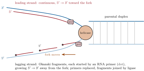
Replication begins at an origin — one on the bacterial chromosome, tens of thousands on a eukaryotic genome — where the helix is opened and two replication forks move away in opposite directions. At each fork: a helicase unwinds the helix (one ATP per base pair), single-strand binding proteins keep the separated strands apart, and a topoisomerase ahead of the fork relieves the twist that unwinding builds up. DNA polymerase adds nucleotides only to the hydroxyl of an existing chain, so that every new strand grows and none can start from nothing: a primase first lays down a short RNA primer that the polymerase extends. On the template read the new strand grows continuously toward the fork: the leading strand. On the other template the new strand must grow away from the fork, in pieces of nucleotides in bacteria and in eukaryotes, the Okazaki fragments, each with its own primer: the lagging strand. A second polymerase replaces the primers with DNA and a ligase seals the fragments into one strand.
Proposition 18.3 (Fidelity)
Base pairing alone would give about one error in ; the polymerase proofreads — a exonuclease activity removes a mispaired nucleotide before the next is added — bringing the error to one in ; and after the fork has passed, a mismatch repair system finds the remaining mispairs, identifies the new strand, and corrects it: one error in to base pairs per replication. A human cell copying pairs makes a handful of mutations each time it divides; a bacterium, one in a thousand divisions.
Example 18.4 (Speed and starting points)
A bacterial fork moves at base pairs a second: two forks copy the of E. coli in , and in rich medium, where the cell divides every twenty, a new round starts before the last has finished. A eukaryotic fork, slowed by chromatin, moves at a second: copying a human chromosome of from one origin would take a month, so the genome is copied from some origins in eight hours, in an order fixed for each cell type.
18.2 The ends
Proposition 18.5 (The end-replication problem and telomerase)
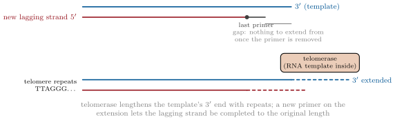
On a linear chromosome the lagging strand cannot be completed to the very end: when the last primer is removed there is no hydroxyl upstream from which to fill the gap, and each replication would shorten the chromosome by nucleotides. Eukaryotes end their chromosomes with telomeres, thousands of copies of a short repeat (TTAGGG in vertebrates) that carry no genes, and germ cells and stem cells carry telomerase, an enzyme with its own RNA template that adds repeats to the end, restoring what replication loses. Most somatic cells lack it: their telomeres shorten at every division, and after some fifty divisions in culture they stop dividing. Bacteria, with circular chromosomes, have no ends.
18.3 The cell cycle
Definition 18.6 (The cell cycle)
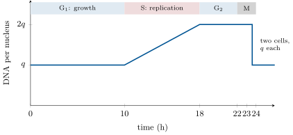
The cell cycle of a eukaryotic cell has four phases. G (gap 1): the cell grows and makes the enzymes of replication; S (synthesis): it replicates its DNA, so that each chromosome becomes two identical sister chromatids joined at the centromere; G: it grows further and checks the copies; M: mitosis, the division of the nucleus, followed by cytokinesis, the division of the cytoplasm. G, S and G together are interphase, in which the chromosomes are extended and active; a cell that stops dividing leaves the cycle from G into a resting state, G. A dividing human cell takes about : G , S , G , M ; a yeast, ; an early frog embryo, thirty minutes with no gaps at all.
Proposition 18.7 (Checkpoints)
The cycle is driven forward by a family of protein kinases, activated in turn by proteins called cyclins whose levels rise and fall through the cycle, and it is held at checkpoints until conditions are met: at the end of G, the cell commits to replicate only if it is large enough, nourished and signalled to divide, and if its DNA is undamaged; at the end of G, it enters mitosis only when replication is complete; in mitosis, the chromatids separate only when every chromosome is attached to the spindle from both sides. Damage arrests the cycle and, if it cannot be repaired, triggers the cell’s death. The molecular machinery of this control belongs to the Year 3 volume; its failure is cancer.
18.4 Mitosis
Definition 18.8 (Mitosis)
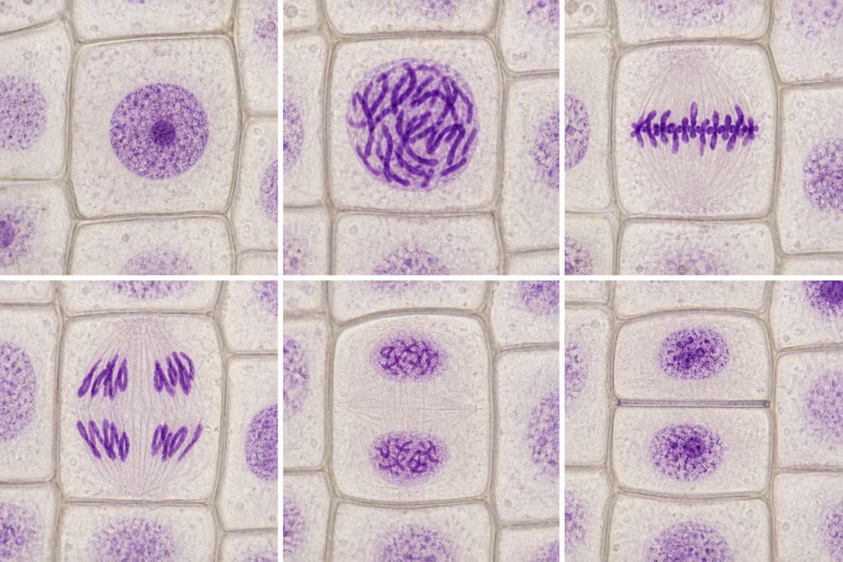
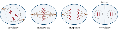
Mitosis distributes the replicated chromosomes so that each daughter nucleus receives one chromatid of every chromosome — the same number and the same genes as the parent. Its stages:
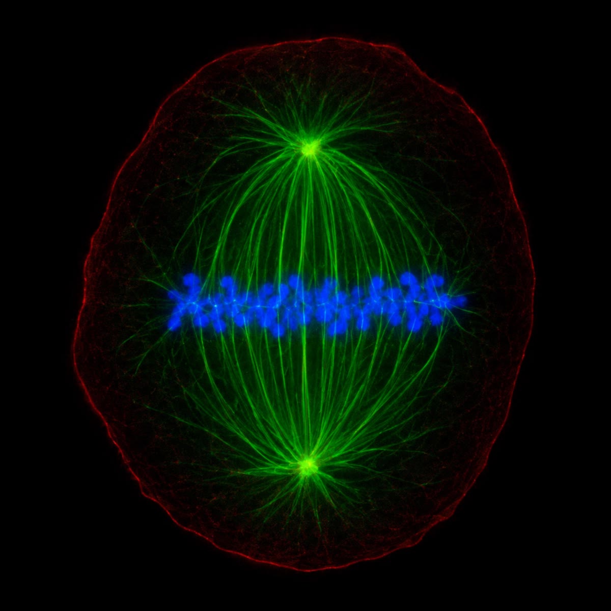
- prophase: the chromosomes condense into visible rods, each of two sister chromatids held together along their length by cohesin proteins; the two centrosomes move apart and the mitotic spindle of microtubules grows between them;
- prometaphase: the nuclear envelope breaks down; spindle microtubules attach to each chromatid’s kinetochore, a protein structure at the centromere, from opposite poles;
- metaphase: the chromosomes are aligned at the equator of the spindle, each under tension from both poles;
- anaphase: the cohesin is cut, the sister chromatids separate and are pulled to opposite poles as the microtubules shorten (anaphase A) and the poles move apart (anaphase B);
- telophase: the chromosomes decondense, and a nuclear envelope re-forms around each set.
Cytokinesis then divides the cytoplasm: in animal cells a contractile ring of actin and myosin pinches the cell in two; in plant cells vesicles from the Golgi fuse at the equator into a cell plate that grows outward into a new wall.
Proposition 18.9 (What mitosis conserves)
Mitosis is a division that conserves the chromosome number: a cell with 2n chromosomes (46 in a human) gives two cells with 2n. The DNA content goes from (after replication) to in each daughter; the number of chromosomes never changes, because a chromosome is counted by its centromere, and each chromatid, once separated, is a chromosome. Every cell of a body is thus genetically identical to the egg it descends from, apart from the mutations that copying has introduced. The division that halves the chromosome number, meiosis, belongs to the Year 2 volume.
Method 18.10 (Reading a cell-cycle experiment)
- Measure the DNA per cell (a dye whose fluorescence is proportional to DNA, cell by cell): cells at are in G, at in G or M, in between in S. The fractions give the durations: a phase’s share of the cycle is its share of the cells.
- Give a pulse of labelled nucleotide (bromodeoxyuridine): only cells in S phase take it up; the labelled fraction is the S fraction, and following the label into mitosis gives the length of G.
- Count mitotic figures in a stained section: the mitotic index, the fraction of cells in M, is the fraction of the cycle that M occupies — for a cycle with a one-hour mitosis.
- Block a step (a drug that depolymerises microtubules arrests cells in metaphase; one that blocks DNA synthesis arrests them in S) and count what accumulates.
Example 18.11 (Turnover)
The epithelium of the small intestine is renewed every four days, the skin every month, the red cells every four months from the marrow, which produces two million a second; a liver cell divides once a year or so and a neuron never. A tumour’s cells have lost the checkpoints: they divide when they should not, tolerate mis-segregated chromosomes, and accumulate the mutations that let them divide faster still. Chemotherapy exploits the difference: drugs that poison the spindle or the polymerase kill the cells that divide most.
18.5 Exercises
Exercise 18.1 ★
Explain why one strand at a fork is copied continuously and the other in fragments.
Solution
Solution of Exercise 18.1.
Polymerases add nucleotides only to a end, so new strands grow ; the two templates are antiparallel, so at a fork one new strand can grow toward the fork continuously while the other must grow away from it, restarting in fragments as more template is exposed.
Exercise 18.2 ★
List the enzymes and proteins at a replication fork and the job of each.
Solution
Solution of Exercise 18.2.
Helicase (unwinds), single-strand binding proteins (hold the strands apart), topoisomerase (relieves twist ahead), primase (RNA primers), DNA polymerase (extends primers, proofreads), a second polymerase (replaces primers), ligase (seals fragments).
Exercise 18.3 ★
From the DNA-content figure, read the DNA content in G, G and after mitosis, and the number of chromosomes in each.
Solution
Solution of Exercise 18.3.
G: , 2n chromosomes of one chromatid. G: , 2n chromosomes of two chromatids. After mitosis: per daughter, 2n chromosomes each.
Exercise 18.4 ★
Order the stages of mitosis and give the defining event of each.
Solution
Solution of Exercise 18.4.
Prophase (condensation, spindle forms), prometaphase (envelope breaks, kinetochores attach), metaphase (alignment at the equator), anaphase (chromatids separate), telophase (envelopes re-form), then cytokinesis.
Exercise 18.5 ★★
Compute the number of Okazaki fragments made in copying the human genome ( pairs, fragments of ), and the number of primers, primer removals and ligations this implies.
Solution
Solution of Exercise 18.5.
Half the genome is lagging-strand synthesis: fragments; as many primers, removals and ligations.
Exercise 18.6 ★★
Compute the error rate per human genome per replication for a polymerase alone (), with proofreading () and with mismatch repair (). How many mutations does each give per division?
Solution
Solution of Exercise 18.6.
; : 640; : about 6 mutations per division.
Exercise 18.7 ★★
A somatic cell loses nucleotides of telomere per division and starts with ; it stops dividing at . How many divisions can it make? Why does a cancer cell need telomerase?
Exercise 18.8 ★★
A tissue has of its cells in mitosis and in S phase; mitosis lasts one hour. Compute the cycle length and the length of S phase.
Solution
Solution of Exercise 18.8.
Cycle ; S .
Exercise 18.9 ★★
A cell has 2n . Give the number of chromosomes, of chromatids and the DNA content (in ) in G, G, metaphase, and each daughter after telophase.
Solution
Solution of Exercise 18.9.
G: 8 chromosomes, 8 chromatids, . G: 8, 16, . Metaphase: 8, 16, . Daughter: 8, 8, .
Exercise 18.10 ★★★
E. coli needs to replicate its chromosome but divides every in rich medium. Explain how, and compute how many origins and forks a cell contains at the moment of division.
Solution
Solution of Exercise 18.10.
New rounds start every at the origin before the previous round ends: replication is overlapping, and a newborn cell already has partly replicated DNA. At division the chromosome finishing its round was started earlier, a second round started earlier has forks halfway, and a third is just beginning: 4 origins and 6 forks in the dividing cell.
Exercise 18.11 ★★★
Colchicine depolymerises microtubules. Predict what happens to a dividing cell treated with it, to its chromosome number if it then re-enters interphase, and why the drug is used to make karyotypes.
Solution
Solution of Exercise 18.11.
No spindle forms: the chromosomes condense but cannot align or separate, and the cell arrests at the spindle checkpoint in a metaphase-like state. If it slips back into interphase without dividing it has 4n chromosomes (tetraploid). Arrested cells with condensed, separate chromosomes are exactly what a karyotype needs: the drug is applied to accumulate them.
Exercise 18.12 ★★★
“Mitosis is a copying of the genome followed by a counting.” Discuss in a paragraph: what replication guarantees, what the spindle guarantees, the checkpoints between them, and what fails in a cell that mis-segregates a chromosome.
Solution
Solution of Exercise 18.12.
Replication guarantees that each chromosome becomes two identical chromatids, to one error in a billion; the spindle guarantees that each daughter receives one of each pair, by attaching every chromosome from both poles and holding anaphase until every one is under tension. The G checkpoint ensures copying is finished before counting begins; the spindle checkpoint ensures counting is set up before separation. A cell that mis-segregates ends with one chromosome too many or too few (aneuploidy): a copy correct to the letter, delivered to the wrong address — which kills most such cells and characterises most cancers.
18.6 Problem: Copying a Human Genome
Problem 18.1
Weekend problem — an S phase of eight hours: forks timed, origins counted, fragments and primers tallied, errors reckoned, and a bacterium compared, ending on the minimum number of origins
A human cell replicates base pairs in an S phase of . A eukaryotic fork moves at ; a bacterial fork at . Okazaki fragments are nucleotides in eukaryotes and in bacteria. Each nucleotide added costs two ATP equivalents (the triphosphate precursor). Error rates per base: for the polymerase alone, with proofreading, with mismatch repair.
Part I — Forks and origins.
- How many base pairs does one fork copy in ?
- An origin sends out two forks. How many base pairs does one origin copy in ?
- Compute the minimum number of origins needed to copy the genome in if all fire at the start.
- Compute the mean spacing of these origins along the DNA, in kilobases and in micrometres.
- In reality origins fire throughout S phase and about are used. Compute the mean spacing and the mean distance one fork actually travels.
- How long would the largest chromosome () take to copy from a single origin at its centre?
- Explain why the number of origins, not the fork speed, is what evolution adjusted to make S phase short.
Part II — Fragments and primers.
- Compute the number of Okazaki fragments made in one S phase.
- Compute the number of RNA primers laid down, including one per leading strand per origin.
- Compute the number of ligations needed.
- Compute the total nucleotides polymerised (both strands of the whole genome).
- Compute the ATP equivalents spent on polymerisation, and the mean rate in ATP per second over S phase.
- Compare with a resting cell’s ATP turnover of about per second: what fraction of the cell’s energy goes to polymerisation?
Part III — Errors.
- Compute the number of errors per S phase at each of the three error rates.
- A gene’s coding sequence is . With the full machinery, what is the probability that a given gene acquires a mutation in one division?
- A person’s cells undergo some divisions in a lifetime. How many mutations in total, at ? Why does the body tolerate this?
- A mutation disables mismatch repair. By what factor does the mutation rate rise, and what does this do to the risk of cancer over a lifetime?
Part IV — The bacterium. E. coli: , one origin, two forks.
- Compute the time to replicate the chromosome.
- In rich medium the cell divides every . How many rounds of replication are under way at once, and how many origins does a newborn cell carry?
- Compute the number of Okazaki fragments per round and per second.
- Compute the errors per round at and the fraction of daughter cells carrying a new mutation.
- A culture of cells per millilitre divides once. How many new mutations appear in a millilitre, and how many hit a given gene of ?
- Explain why antibiotic resistance can be found in almost any large culture of a sensitive strain.
- Compare the two organisms: base pairs, origins, fork speed, S-phase duration, and the design that lets the larger genome be copied in a comparable time.
- State the result: the minimum number of origins for a human S phase of , and the real number and spacing.
Solution
Solution of Problem 18.1.
1. . 2. . 3. origins. 4. , i.e. of DNA between origins. 5. spacing; each fork travels about , half an hour’s work. 6. per fork at : s, 29 days. 7. The fork speed is limited by the chemistry and by chromatin (a twentyfold slowing relative to bacteria); origins can be multiplied without limit, so S phase is shortened by adding starting points, not by racing the forks. 8. Lagging synthesis covers half the genome, nucleotides: fragments (each origin’s two forks each have a lagging strand, and the halves add to one genome’s worth). 9. primers. 10. About ligations (one per fragment) plus one per meeting of forks. 11. nucleotides. 12. ATP; over , per second. 13. About of the cell’s ATP turnover: polymerisation is cheap; what is expensive is making the nucleotides and the histones. 14. , 640 and 6.4 errors. 15. : one gene in seven hundred thousand per division. 16. mutations — every possible single change of the genome many times over, scattered among cells: almost all fall in non-coding DNA or in cells that are shed, and a mutated cell is one among billions; only a few combinations in one cell matter. 17. A hundredfold (): the sequence of mutations that makes a cancer, which takes decades to accumulate at , accumulates far sooner — inherited defects in mismatch repair cause colon cancer in early adulthood. 18. pairs per fork at : , . 19. About two rounds overlapping; a newborn cell carries two origins (each already replicated once) and forks halfway along: its chromosome is partly replicated at birth. 20. fragments per round, about 1.3 per second. 21. errors per round: about one daughter in 200 carries a new mutation. 22. new mutations per millilitre; a given gene is hit times. 23. With a thousand independent mutations in any given gene per millilitre per division, every possible single change — including the ones that confer resistance — is already present in a large culture before the antibiotic is applied; the drug selects, it does not create. 24. Human: pairs, origins, , . Bacterium: pairs, one origin, , . A genome a thousand times larger, copied by forks twenty times slower, in only twelve times the time: the difference is the tens of thousands of origins working in parallel. 25. About origins at minimum, if all fired at once; in reality some , one every , firing in succession so that each fork travels about .