University Biology — Year 1 · Bachelor Year 1
11Nucleotides and Nucleic Acids
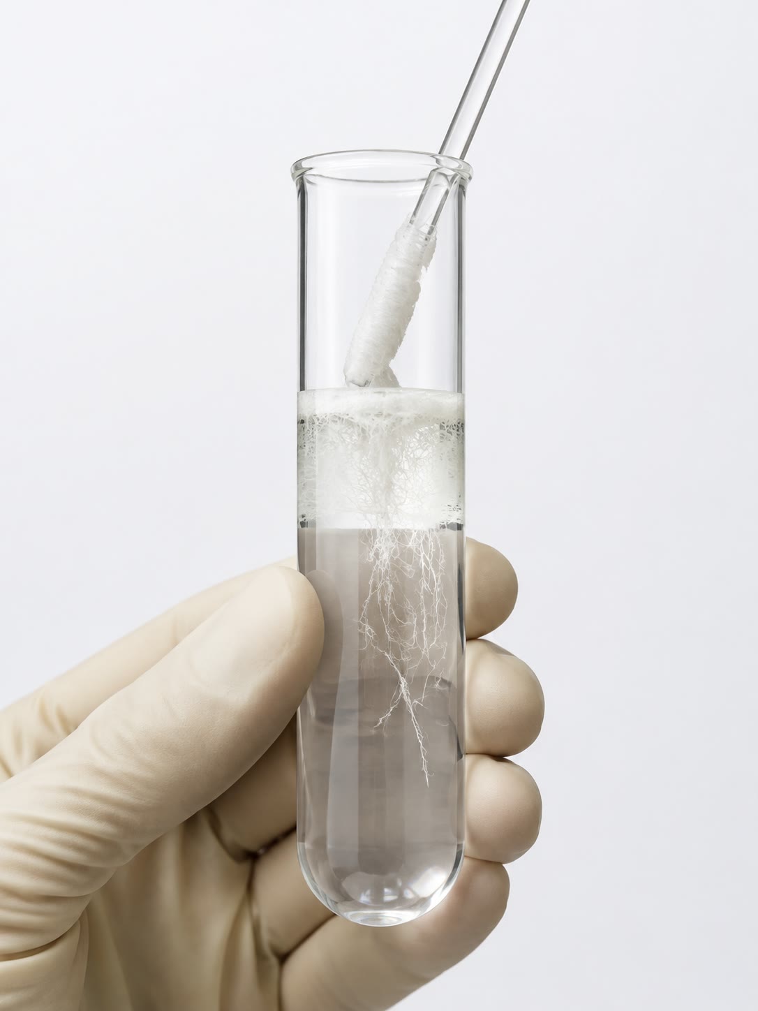
Pour cold ethanol onto a solution of broken cells and a white cloud of fibres appears that can be wound onto a glass rod: DNA, the molecule that carries every instruction of the organism, visible to the naked eye. Two metres of it are folded into every human nucleus; the whole of it is written in an alphabet of four letters, read in one direction, and copied by separating two strands that fit each other like a hand and its glove. This chapter describes the nucleotides — letters, energy currency and coenzymes at once — the chains they form, the double helix and the evidence for it, what happens when it is melted and re-annealed, and the RNAs that carry its message into the cell.
11.1 Nucleotides
Definition 11.1 (Nucleotide)
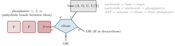
A nucleotide is made of three parts: a nitrogenous base — a purine (adenine A, guanine G: two fused rings) or a pyrimidine (cytosine C, thymine T, uracil U: one ring); a five-carbon sugar, ribose in RNA or 2-deoxyribose in DNA (lacking the hydroxyl on carbon ); and one to three phosphate groups on the sugar’s carbon . The base is attached to carbon ; the sugar carbons are numbered with primes to distinguish them from the base’s. Base plus sugar is a nucleoside (adenosine, guanosine, cytidine, thymidine, uridine); with phosphates it is a nucleotide (AMP, ADP, ATP and their kin).
Proposition 11.2 (Nucleotides do three jobs)
Beyond being the monomers of DNA and RNA, nucleotides are the cell’s energy currency — ATP and GTP, whose phosphoanhydride bonds (Chapter 8) pay for synthesis, transport and movement; its coenzymes — , and FAD, which carry electrons in respiration and photosynthesis (Chapter 15), and coenzyme A, which carries acyl groups; and its messengers — cyclic AMP, which relays hormone signals inside cells. All of them are adenine nucleotides or built on them: the same molecule that spells the message powers its execution.
11.2 The polynucleotide chain
Definition 11.3 (Phosphodiester bond, polarity)
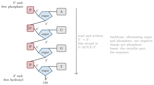
Nucleotides are joined into a polynucleotide by phosphodiester bonds: the phosphate on carbon of one nucleotide is linked to the hydroxyl of the next. The chain therefore has a sugar–phosphate backbone, uniform and negatively charged (one charge per phosphate), from which the bases project as a sequence; and it has a direction: a end bearing a free phosphate and a end bearing a free hydroxyl. Sequences are written and read , the direction in which chains are synthesised. DNA (deoxyribonucleic acid) uses deoxyribose and the bases A, G, C, T; RNA (ribonucleic acid) uses ribose and A, G, C, U.
Example 11.4 (RNA is fragile, DNA is durable)
The hydroxyl of ribose can attack the neighbouring phosphodiester bond: in mild alkali RNA is hydrolysed to nucleotides within hours, while DNA, lacking that hydroxyl, survives boiling in alkali and has been recovered intact from bones tens of thousands of years old. Uracil in RNA is thymine without a methyl group; DNA uses thymine so that cytosine, which slowly loses its amino group to become uracil, can be recognised as damaged and repaired — a uracil in DNA is always a mistake.
11.3 The double helix
Proposition 11.5 (Chargaff’s rules)
In the DNA of any organism the amount of adenine equals that of thymine and the amount of guanine that of cytosine (, , hence purines pyrimidines), while the ratio varies from species to species (from to ).
Evidence. Chargaff (1950) hydrolysed DNA from many organisms and separated the bases by chromatography, measuring each by its ultraviolet absorption. The equalities held within experimental error for every DNA; the content did not, and was the same in every tissue of one organism. ∎
Theorem 11.6 (The Watson–Crick double helix)
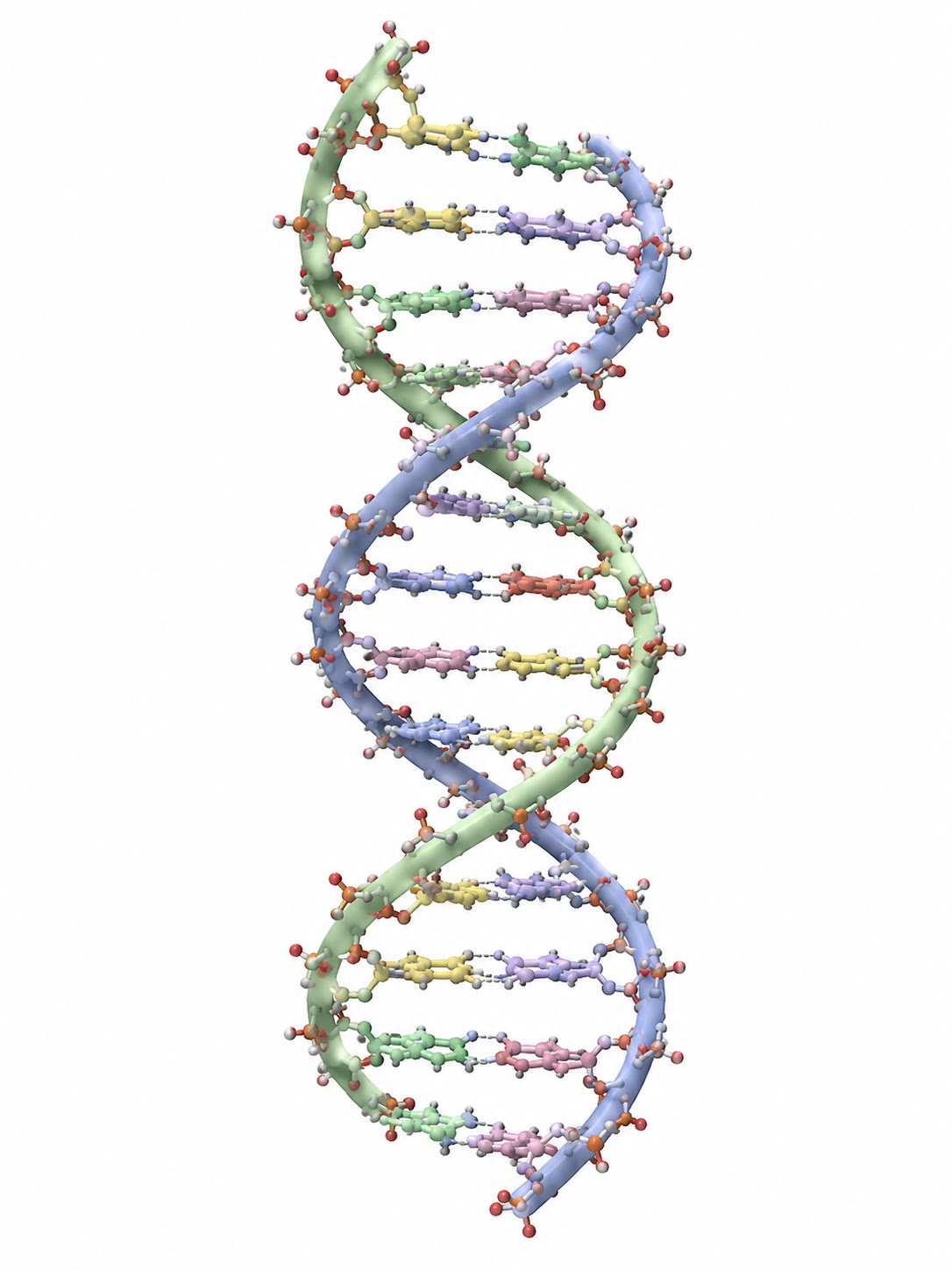
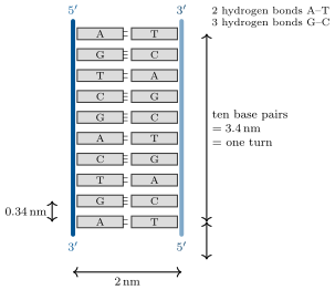
DNA is two polynucleotide chains wound around a common axis into a right-handed double helix: the sugar–phosphate backbones outside, the bases inside, flat and stacked perpendicular to the axis. The chains are antiparallel (one runs upward, the other downward) and complementary: opposite every A is a T, held by two hydrogen bonds, and opposite every G a C, held by three; each such base pair is the same width, so the helix is uniform whatever the sequence. In the common B form the helix is across, rises per base pair and per turn of ten pairs, and shows a wide major groove and a narrow minor groove in which the edges of the bases are exposed to proteins.
Evidence. Franklin’s X-ray diffraction pictures of DNA fibres (1952) showed the cross of a helix with a repeat of along the axis, a strong reflection at (stacked flat units), and a diameter of ; the phosphates, the heaviest atoms, had to be outside. Watson and Crick (1953) found that a purine paired with a pyrimidine — and only A with T and G with C — gives pairs of equal width fitting the diameter, explaining Chargaff’s rules, and that two antiparallel chains so paired build a regular helix with the fibre’s dimensions. The model predicted that each chain is a template for the other; Meselson and Stahl’s demonstration of semi-conservative replication (recalled from the High School volume, and in Chapter 18) confirmed it. ∎
Example 11.7 (Reading a strand)
If one strand reads -ATGGCATTC-, its partner, written , is -GAATGCCAT-: complement each base, then reverse the order. Nine pairs, of helix, twenty-two hydrogen bonds (five G–C, four A–T). A human chromosome of pairs is long in this form and must be folded a hundred thousand times to fit a nucleus (Chapter 17).
Proposition 11.8 (What holds the helix together)
The hydrogen bonds between paired bases give specificity; the stacking of the flat bases on one another — van der Waals contacts and the hydrophobic effect that keeps the bases out of water — gives most of the stability; the negatively charged backbones repel each other, and the helix is stable only in the presence of cations (the physiological , , or the basic proteins of chromatin) that screen the charge. A G–C pair, with three hydrogen bonds and stronger stacking, holds better than an A–T pair.
11.4 Melting and hybridisation
Definition 11.9 (Denaturation, melting temperature)
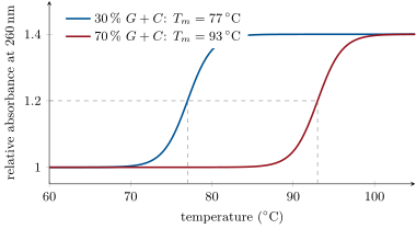
Heating, or extreme pH, separates the two strands of DNA: denaturation (melting), a cooperative transition over a few degrees, whose midpoint is the melting temperature . It is followed by the hyperchromic effect: single strands absorb about more ultraviolet light at than the same bases stacked in a helix. rises with the content ( per percent), with the salt concentration, and with length; in salt a long DNA of melts near . Cooled slowly, the strands find their complements and re-form the helix: renaturation, or hybridisation when the strands come from different sources.
Method 11.10 (Measuring nucleic acids by absorbance)
- Bases absorb ultraviolet light with a maximum at . In a cell, an absorbance of corresponds to of double-stranded DNA ( of RNA, of single-stranded DNA).
- The ratio is for pure DNA and for pure RNA; proteins absorb at and lower it.
- To follow melting, record while heating slowly; the curve’s midpoint is and its steepness a measure of the homogeneity of the sample.
- To detect a sequence, denature the sample and add a labelled single strand complementary to it (a probe); it hybridises only where its complement is, and the label marks the place — on a gel, on a chromosome, in a tissue.
Example 11.11 (A probe finds a gene)
A twenty-nucleotide probe complementary to one gene, at below its own , binds its exact complement and nothing else in three billion base pairs: a single mismatch in twenty lowers by several degrees and the mismatched hybrid melts. The specificity of base pairing, multiplied over twenty positions, is what every DNA test, from paternity to pathogen detection, relies on.
11.5 RNA
Definition 11.12 (The RNAs)
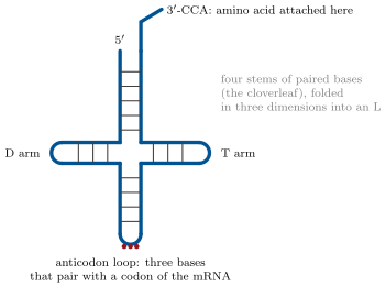
RNA is usually single-stranded, and folds back on itself wherever short complementary stretches allow, into hairpins, loops and bulges — a secondary structure — that then packs into a shape. Three classes carry the flow of genetic information (Chapter 19): messenger RNA (mRNA), a copy of a gene that the ribosome reads; transfer RNA (tRNA), some nucleotides folded into a cloverleaf, carrying an amino acid at its end and an anticodon that pairs with the message; ribosomal RNA (rRNA), the structural and catalytic core of the ribosome, four fifths of the RNA of a cell. Many other RNAs regulate, splice, and guide (the Year 3 volume).
Remark 11.13 (RNA can be a catalyst)
Some RNAs are enzymes (ribozymes): the peptide bond of every protein is formed by the ribosomal RNA, not by a protein, and some RNAs cut and splice themselves. A molecule that both carries information and catalyses reactions is what a first living system may have been made of; the DNA–protein world would then be a later division of labour, with RNA kept at its centre.
11.6 Exercises
Exercise 11.1 ★
Name the three parts of a nucleotide and the two differences between a ribonucleotide and a deoxyribonucleotide.
Solution
Solution of Exercise 11.1.
A nitrogenous base, a pentose, one to three phosphates. RNA: ribose (with a hydroxyl) and uracil; DNA: -deoxyribose and thymine.
Exercise 11.2 ★
Write the complementary strand of -GGATCCTA- in the direction, and count its hydrogen bonds.
Solution
Solution of Exercise 11.2.
-TAGGATCC-. Pairs: G–C, G–C, A–T, T–A, C–G, C–G, T–A, A–T: four G–C (12 bonds) and four A–T (8): 20 hydrogen bonds.
Exercise 11.3 ★
A DNA contains adenine. Give the percentages of T, G and C, and the content.
Solution
Solution of Exercise 11.3.
T ; G and C share the remaining : each; .
Exercise 11.4 ★
From the melting figure, what is the of a DNA of in the same conditions, and by how much does the absorbance rise on melting?
Solution
Solution of Exercise 11.4.
Between the two curves, at per percent: about . The absorbance rises by .
Exercise 11.5 ★★
Compute the length of the E. coli chromosome ( base pairs) in millimetres, its number of turns, and the factor by which it must be compacted to fit in a cell of .
Solution
Solution of Exercise 11.5.
; turns; -fold in length (the chromosome is folded into loops and supercoils).
Exercise 11.6 ★★
Explain why the two strands of DNA must be antiparallel, using the geometry of the sugar–phosphate backbone and the requirement that every base pair have the same width.
Solution
Solution of Exercise 11.6.
The bases pair with their edges facing, and the sugar of each is attached at the same side of the pair only if the two backbones run in opposite directions; with parallel strands the two sugars would sit on opposite edges and the width of the pair, and the position of the backbone, would vary with the sequence. Antiparallel strands with purine–pyrimidine pairs give the constant that the fibre pattern requires.
Exercise 11.7 ★★
A solution of DNA has in a cell and . Compute the concentration and comment on the purity. How many base pairs are in , if the mean mass of a pair is ?
Solution
Solution of Exercise 11.7.
. : essentially pure DNA (1.8), with perhaps a trace of protein. In : base pairs.
Exercise 11.8 ★★
Explain the hyperchromic effect and why the melting curve of a pure DNA is steep while that of a mixture of DNAs from many species is spread out.
Solution
Solution of Exercise 11.8.
Stacked bases shield one another and absorb less; separating the strands exposes each base to the light and the absorbance rises . A pure DNA melts within a few degrees of its single , cooperatively; a mixture contains DNAs of many contents, each with its own , so the sum of their curves is spread over tens of degrees.
Exercise 11.9 ★★
A twenty-nucleotide probe has with its exact complement; one mismatch lowers by about . At which temperature should hybridisation be done to detect the exact sequence and reject single mismatches? What happens at ?
Solution
Solution of Exercise 11.9.
Just below and above , say : the perfect hybrid holds, the mismatched one melts. At hybrids with up to three mismatches are stable and the probe also lights up related sequences.
Exercise 11.10 ★★★
Chargaff’s rules hold for double-stranded DNA but not for RNA or for the DNA of certain small viruses. Explain what each exception tells about the structure of those nucleic acids.
Solution
Solution of Exercise 11.10.
The rules follow from base pairing between two complementary strands. RNA, single-stranded, has no partner to constrain its composition, so in general; the small viruses in question have single-stranded DNA genomes, which likewise need not obey the rules — and their failure to do so was one of the first hints that they were single-stranded.
Exercise 11.11 ★★★
Two hydrogen bonds hold an A–T pair, yet a helix of a thousand A–T pairs is stable at while a single pair does not form at all. Explain with the ideas of Chapter 8 (weak bonds, cooperativity, stacking), and say what this implies for the fidelity of a probe.
Solution
Solution of Exercise 11.11.
Each pair alone is worth a few kilojoules per mole, less than thermal agitation, and forms and breaks in microseconds. In a helix the pairs form together: each pair stacks on the last and the first pairs nucleate the rest, so the energies add and the probability that all break at once is negligible — cooperativity. Stacking contributes as much as the hydrogen bonds. For a probe this means that the stability grows with length and falls sharply with each mismatch, which breaks the cooperative run: specificity is a property of the whole stretch.
Exercise 11.12 ★★★
“The structure of DNA is the explanation of heredity.” Discuss in a paragraph: what the double helix explains immediately (copying, mutation as a change of sequence), what it does not explain by itself (how the sequence becomes a protein), and why Watson and Crick’s model was accepted before any biochemical test of it.
Solution
Solution of Exercise 11.12.
The helix explains copying at once: each strand specifies the other, so separating them and pairing new nucleotides on each gives two identical molecules; a mutation is a wrong base fixed in one copy and faithfully copied thereafter. It does not, by itself, say how a sequence of four bases specifies a protein — that took a decade (code, mRNA, tRNA, ribosome). The model was accepted because it fitted every measurement (Chargaff, the X-ray dimensions, the density) with nothing left over, and because its explanation of heredity was so economical that the alternative — a coincidence — was implausible; Meselson and Stahl then confirmed the copying it predicted.
11.7 Problem: The Genome of a Virus
Problem 11.1
Weekend problem — the DNA of phage measured, weighed, counted, melted and packed into its capsid, ending on its melting temperature and its contour length
The genome of bacteriophage is one linear double-stranded DNA of base pairs with a content of . It is packed into an icosahedral capsid of internal diameter. Take per base pair, for the helix diameter, per base pair, and in degrees Celsius, with in and the length in base pairs.
Part I — Measured and weighed.
- Compute the contour length of the molecule in micrometres.
- Compute the number of turns of the helix.
- Compute its molar mass in daltons.
- Compute the mass of one molecule in grams.
- Compute the number of phosphate groups and hence the net charge of the molecule.
- Compute the number of nucleotides of each base (A, T, G, C).
- Compute the total number of hydrogen bonds holding the two strands.
Part II — Melted.
- Compute in sodium.
- Compute in sodium, and explain why salt stabilises the helix.
- The genome contains a region of pairs at and one at . Compute the of each region in sodium and describe the shape of the melting curve of the whole molecule.
- A solution of DNA has before melting. What is its concentration, and what will be after complete melting?
- How many molecules are in of that solution?
- The two ends of the genome are single-stranded overhangs of 12 nucleotides, complementary to each other, by which the molecule circularises in the host. Compute their (use ) and say why the circle nevertheless holds at inside the cell.
Part III — Packed.
- Compute the volume of the DNA as a cylinder.
- Compute the internal volume of the capsid.
- What fraction of the capsid does the DNA fill? Compare with the closest packing of parallel cylinders ().
- The DNA carries negative charges in a sphere of . Explain what must accompany the DNA into the capsid, and why the packed DNA exerts a pressure on the shell (tens of atmospheres) that helps inject it into a bacterium.
- The molecule must be bent into loops of radius comparable to the capsid’s. DNA resists bending below a radius of about . Comment.
Part IV — Read.
- One strand of the genome starts -GGGCGGCGACCT-. Write the complementary strand .
- If a gene of base pairs is transcribed into an mRNA, what is the mRNA’s length in nucleotides and, at per nucleotide, its mass?
- The genome has genes in pairs. What fraction of the genome is coding if the average gene is pairs? Compare with a human genome ().
- The genome is replicated in at by the host’s enzymes. Compute the rate in base pairs per second if replication starts at one origin and proceeds in both directions.
- How many hydrogen bonds are broken per second at the two forks during replication (use the mean per pair from question 7)?
- A mutation changes one G–C pair to A–T. By how much does the of the whole molecule change? Could melting detect it?
- State the result: the melting temperature of DNA in sodium and its contour length, and the compaction that fits it into the capsid.
Solution
Solution of Problem 11.1.
1. . 2. turns. 3. . 4. . 5. Two per pair: phosphates, net charge . 6. : ; . 7. . 8. . 9. . Cations screen the repulsion between the two negatively charged backbones; with less salt the strands repel more and separate at a lower temperature. 10. : ; : . The whole molecule melts in steps: A–T-rich regions open first, the G–C-rich ones last, so the curve is a broad rise with shoulders rather than a single sharp step. 11. ; after melting . 12. molecules. 13. (the formula is rough for such short stretches). At the ends pair but would breathe open; in the cell an enzyme (ligase) seals the two nicks covalently, and the circle no longer depends on the twelve pairs. 14. . 15. . 16. : two thirds of the closest possible packing of cylinders — the DNA is wound in nearly crystalline coils. 17. Counter-ions (polyamines, ) must neutralise most of the charge or the repulsion could not be overcome; the remaining repulsion and the bending energy stored in the tight coils push outward on the shell, and when the tail opens this pressure drives the first part of the genome into the bacterium. 18. The loops in a capsid have radii of , below the at which DNA bends freely: bending it costs energy, paid by the packaging motor (ATP) and stored as the pressure of question 17. 19. -AGGTCGCCGCCC-. 20. nucleotides; . 21. pairs, coding — a compact genome; the human genome is sixty times less dense in genes. 22. Two forks, each copying pairs in : pairs per second per fork (the host’s polymerase is capable of ; the time is set by the whole cycle, not the polymerase). 23. bonds per pair; two forks at 20 pairs per second: 100 hydrogen bonds per second — and as many re-formed behind them. 24. One pair in changes by and by : undetectable by melting the whole molecule; only a short probe spanning the site would show it. 25. in sodium; contour length , three hundred times the capsid’s diameter, packed into it at of its volume.