University Biology — Year 1 · Bachelor Year 1
13Enzymes and Biochemical Catalysis
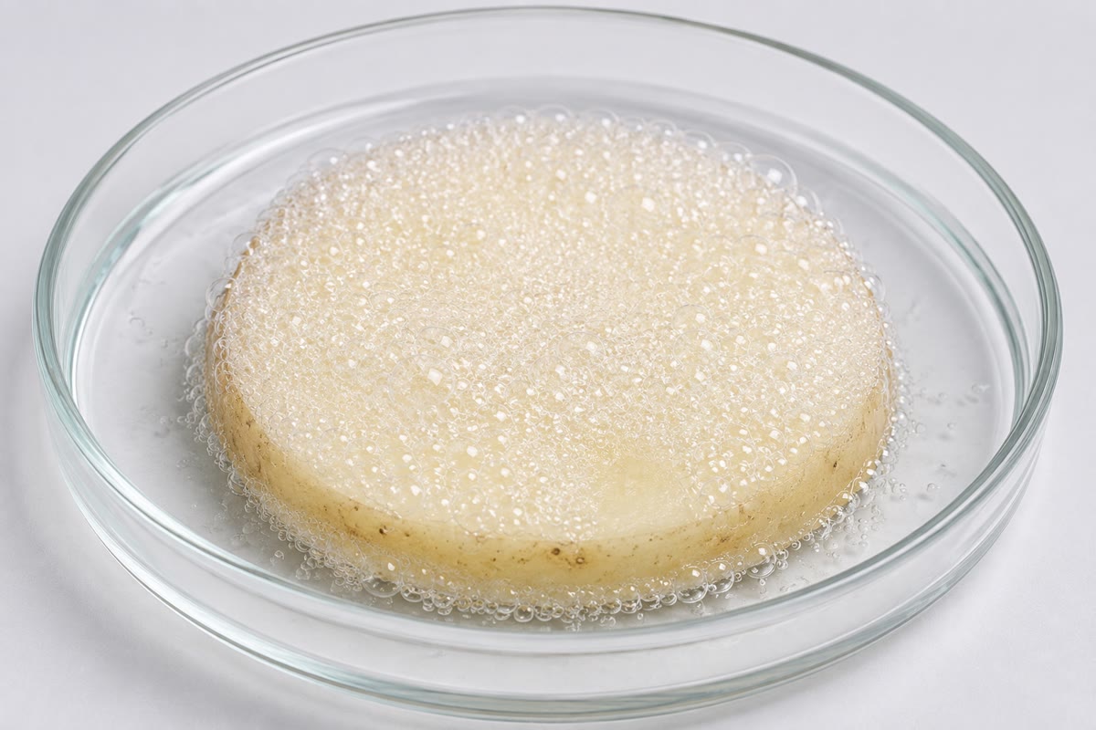
Pour hydrogen peroxide on a cut potato and it froths: a single molecule of catalase in the potato’s cells splits forty million molecules of peroxide every second, a reaction that, left to itself, would take years. The peroxide was going to decompose anyway — the reaction is downhill — but not in any useful time. Every reaction of the cell is like this: thermodynamics says which way it can go, and an enzyme says whether it goes now. This chapter describes what an enzyme does to a reaction, the kinetics by which enzymes are measured and compared, the ways they are inhibited, and the ways the cell switches them on and off.
13.1 Catalysis
Definition 13.1 (Enzyme, substrate, active site)
An enzyme is a protein (rarely an RNA) that catalyses a reaction: it increases the rate without being consumed and without changing the equilibrium. The molecules it acts on are its substrates; they bind in the active site, a cleft a few residues line, where the chemistry happens. Enzymes are specific — for one substrate or a family, for one bond, for one stereoisomer — and they are named for their substrate and reaction with the suffix -ase (lactase, DNA polymerase, succinate dehydrogenase), in six classes: oxidoreductases, transferases, hydrolases, lyases, isomerases, ligases. Many need a non-protein cofactor: a metal ion (Zn, Mg, Fe), or an organic coenzyme — , FAD, coenzyme A, most of them made from vitamins, which is what vitamins are for.
Proposition 13.2 (What an enzyme changes and what it does not)
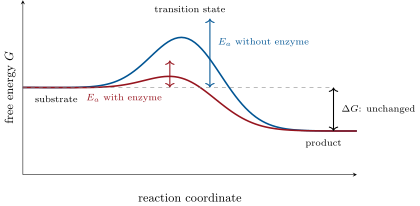
A reaction passes through a transition state, a strained arrangement of the atoms higher in free energy than reactants or products by the activation energy ; only the molecules that thermal agitation carries over this barrier react, in a fraction proportional to . An enzyme binds the transition state more tightly than the substrate — its active site is complementary to the strained form — and so lowers : lowering it by (the worth of a few hydrogen bonds) at multiplies the rate by . It does not change or the equilibrium constant, which depend only on the reactants and products: an enzyme speeds the forward and backward reactions equally and brings the system to the same equilibrium faster.
Proposition 13.3 (How the active site works)
An active site accelerates its reaction by several means at once: it binds and orients the substrates so that the reacting groups meet in the right geometry, replacing a rare collision by a certain one; it strains the substrate toward the transition state (induced fit: the enzyme closes around the substrate, and the fit is best for the transition state); its side chains act as acids and bases, handing protons to and from the substrate at the right moment (histidine, with its p near 7, does this in many enzymes); some form a transient covalent bond with the substrate (serine proteases); and its metal ions polarise bonds and stabilise charges. Water is largely excluded from the site, so that charges and hydrogen bonds act at full strength.
Example 13.4 (Three enzymes)
Lysozyme, in tears and egg white, strains a sugar ring of the bacterial wall into the shape of the transition state and cuts the chain: -fold acceleration. Carbonic anhydrase, in red cells, holds a zinc ion that turns water into a hydroxide poised to attack : a million molecules a second, the fastest enzyme after catalase. Chymotrypsin, in the pancreatic juice, uses a serine made reactive by a histidine to cut proteins after aromatic residues: a hundred per second, with a covalent intermediate.
13.2 Enzyme kinetics
Definition 13.5 (Initial rate, saturation)
The rate of an enzyme reaction is the amount of product formed per unit time (, or units of enzyme: ). It is measured as the initial rate , before the substrate is depleted or the product accumulates. At a fixed enzyme concentration rises with the substrate concentration and then saturates at a maximum : the enzyme is fully occupied and can go no faster.
Theorem 13.6 (Michaelis–Menten)
For an enzyme that binds its substrate reversibly and converts it,
the initial rate at substrate concentration is
, the Michaelis constant, is the substrate concentration at which the rate is half its maximum, and measures how much substrate the enzyme needs; , the turnover number, is the number of substrate molecules one enzyme converts per second when saturated; the ratio is the catalytic efficiency, the rate constant at low substrate, bounded above by the rate at which substrate can reach the enzyme by diffusion, about – .
Proof. Assume a steady state in which is formed as fast as it disappears (valid after the first milliseconds, while ): , so with as defined. Conservation gives ; substituting, , hence . The rate is , which gives the formula; at , , and at , . ∎
![Left: the Michaelis–Menten hyperbola for K_m = 2\, mmol/ L and V_ = 1\, µ mol/ min, with the six measured points of the weekend problem. Right: the same data as a Lineweaver–Burk plot, 1/v_0 against 1/[S]: a straight line whose intercepts give V_ and K_m.](/images/onecourse/chapters/biology-3/b1-enzymes/fig-fcc855eda471.svg)
![Left: the Michaelis–Menten hyperbola for K_m = 2\, mmol/ L and V_ = 1\, µ mol/ min, with the six measured points of the weekend problem. Right: the same data as a Lineweaver–Burk plot, 1/v_0 against 1/[S]: a straight line whose intercepts give V_ and K_m.](/images/onecourse/chapters/biology-3/b1-enzymes/fig-128694e7a7f3.svg)
Method 13.7 (Measuring and )
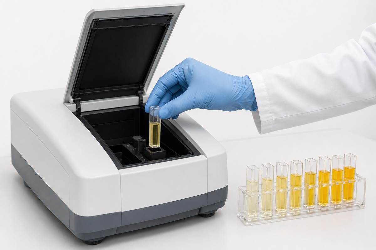
- Prepare a series of substrate concentrations spanning to , with the same enzyme concentration; follow the product (colour, absorbance, gas) for the first minute and take the slope as .
- Plot against : the hyperbola gives a rough (the plateau) and (the concentration at half of it).
- For precision, invert: (Lineweaver–Burk). The line’s intercept on the axis is , on the axis , and its slope .
- Divide by the enzyme concentration to get ; divide by for the efficiency. Repeat with an inhibitor to see which parameter it changes.
Example 13.8 (Enzymes compared)
Catalase and carbonic anhydrase work at the diffusion limit: every collision with substrate is productive. Chymotrypsin is slow but needs to be — it cuts a peptide bond, a far harder job than splitting peroxide.
13.3 Inhibition
Definition 13.9 (Inhibitors)
An inhibitor lowers the rate of an enzyme. Irreversible inhibitors bind covalently and destroy the enzyme’s activity for good (nerve agents on acetylcholinesterase, penicillin on the enzyme that cross-links the bacterial wall, aspirin on the enzyme that makes prostaglandins). Reversible inhibitors bind by weak bonds and can be washed away; by where they bind:
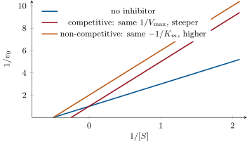
- competitive: the inhibitor resembles the substrate and occupies the active site; it raises the apparent (by the factor ) and leaves unchanged — enough substrate displaces it;
- non-competitive: the inhibitor binds elsewhere and spoils the catalysis whether or not substrate is bound; it lowers and leaves unchanged — no amount of substrate overcomes it;
- uncompetitive: the inhibitor binds only the complex; both and fall.
, the dissociation constant of the inhibitor, measures its potency: the lower, the stronger.
Example 13.10 (Competition as medicine)
Methanol is harmless until the liver’s alcohol dehydrogenase turns it into formaldehyde and formic acid, which blind and kill. The treatment is ethanol: a competing substrate with a lower , which keeps the enzyme busy while the methanol is excreted unchanged. Statins are competitive inhibitors, resembling the transition state, of the enzyme that commits carbon to cholesterol; sulfonamides resemble the substrate of a bacterial enzyme that makes folate, which humans do not make and so do not miss.
13.4 Regulation
Definition 13.11 (Allosteric enzymes)
An allosteric enzyme has several subunits and, besides its active sites, regulatory sites where effectors bind: activators shift it toward its active conformation, inhibitors toward the inactive one. Its rate against is sigmoid, not hyperbolic (cooperativity among the active sites, as for haemoglobin, Chapter 12), so that a small change of substrate near the steep part changes the rate greatly; effectors shift the curve sideways (changing the substrate concentration needed) or up and down (changing the maximal rate). Allosteric enzymes stand at the branch points of metabolism, and the effectors are the pathway’s own products and the cell’s energy signals (ATP, ADP, AMP).
![An allosteric enzyme. The sigmoid curve makes the rate sensitive to substrate near the midpoint; an activator shifts it left (more active at a given [S]), an inhibitor right. Near [S] = 3 the rate can swing from a tenth to nine tenths of maximum.](/images/onecourse/chapters/biology-3/b1-enzymes/fig-0bb8ee6cbc43.svg)
Proposition 13.12 (Four ways the cell controls an enzyme)
- Feedback inhibition: the end product of a pathway is an allosteric inhibitor of its first committed enzyme, so that the pathway runs only as fast as the product is used (isoleucine on threonine deaminase; ATP on phosphofructokinase, Chapter 15). Response time: milliseconds.
- Covalent modification: a kinase attaches a phosphate to a serine, threonine or tyrosine of the enzyme, a phosphatase removes it, and the two forms differ in activity (glycogen phosphorylase is switched on by phosphorylation, glycogen synthase off, by the same hormonal signal). Seconds to minutes; reversible; amplifiable in cascades.
- Proteolytic activation: some enzymes are made as inactive precursors (zymogens: trypsinogen, pepsinogen, the clotting factors) and switched on by cutting off a peptide, once and for all, where and when they are wanted.
- Amount: the cell makes more or less of the enzyme by controlling its gene (Chapter 20) and degrades it faster or slower. Minutes to hours.
Example 13.13 (Isoenzymes)
Lactate dehydrogenase exists in five forms, tetramers of two subunit types in all combinations; the heart’s form has a low for lactate and is inhibited by pyruvate (it oxidises lactate to feed the Krebs cycle), the muscle’s form has a high and tolerates pyruvate (it makes lactate in a sprint). The pattern of forms in the blood reveals which organ has been damaged — a heart attack releases the heart’s.
13.5 Temperature and pH
Proposition 13.14 (Enzymes and their environment)
The rate of an enzyme reaction roughly doubles for every () as thermal energy carries more molecules over the barrier, until the enzyme begins to unfold ( for most; for the enzymes of hot-spring bacteria) and the rate collapses. Each enzyme has a pH optimum, where the ionisation of its catalytic residues and of its substrate is right: pepsin, in the stomach, near pH 2; trypsin, in the intestine, near 8; most intracellular enzymes near 7. Away from the optimum the rate falls, and far from it the protein denatures. Temperature and pH act on the protein; the cell keeps both within the range where its thousand enzymes all work.
Example 13.15 (A fever and a hot spring)
A fever of speeds every reaction of the body by a quarter and begins to unfold the most fragile proteins; at the brain’s fail. The DNA polymerase of a bacterium from a spring survives , which is why it can be cycled through the melting of DNA thousands of times and became the enzyme of the polymerase chain reaction (the Year 3 volume): thermostability is a property of the sequence, not of the chemistry catalysed.
13.6 Exercises
Exercise 13.1 ★
What does an enzyme change in a reaction, and what does it leave unchanged? Illustrate with the energy profile.
Solution
Solution of Exercise 13.1.
It lowers the activation energy (the height of the transition state), and so the rate, in both directions; it leaves , the equilibrium constant and the position of equilibrium unchanged. On the profile the peak is lowered, the two ends are not.
Exercise 13.2 ★
Define , , and , with units.
Exercise 13.3 ★
From the Lineweaver–Burk figure of the inhibitors, read and for the uninhibited enzyme and for each inhibitor.
Solution
Solution of Exercise 13.3.
No inhibitor: intercept 1, so ; intercept , so . Competitive: , intercept , . Non-competitive: intercept 2, ; intercept , .
Exercise 13.4 ★
Name the four ways a cell regulates an enzyme’s activity and give the timescale of each.
Solution
Solution of Exercise 13.4.
Allosteric (feedback) regulation, milliseconds; covalent modification (phosphorylation), seconds to minutes; proteolytic activation of a zymogen, once, on demand; control of the amount by gene expression and degradation, minutes to hours.
Exercise 13.5 ★★
An enzyme has and . Compute at , , and . At what is ?
Solution
Solution of Exercise 13.5.
: 8.3, 25, 45.5 and . gives .
Exercise 13.6 ★★
Lowering by at multiplies the rate by what factor? By how much must fall to gain a factor ?
Solution
Solution of Exercise 13.6.
. For : .
Exercise 13.7 ★★
of carbonic anhydrase in gives per minute. Compute and, with , the efficiency. Compare with the diffusion limit.
Exercise 13.8 ★★
A competitive inhibitor at doubles the apparent . Compute . What substrate concentration restores the rate to of with and without the inhibitor, if ?
Exercise 13.9 ★★
Explain why feedback inhibition acts on the first committed step of a pathway rather than the last, and why the inhibitor is usually the end product rather than an intermediate.
Solution
Solution of Exercise 13.9.
Inhibiting the first committed step stops the whole pathway at once and wastes no intermediates, which would otherwise accumulate; the end product is the signal that matters — its abundance is exactly the information that the pathway is no longer needed — whereas an intermediate’s level says nothing about demand.
Exercise 13.10 ★★★
Trypsinogen is activated in the intestine, not in the pancreas that makes it; a small fraction activated early destroys the pancreas. Explain the logic of zymogens, the role of the enzyme that activates trypsinogen, and why the pancreas also makes a trypsin inhibitor.
Solution
Solution of Exercise 13.10.
A protease active in the cell that makes it would digest that cell; made as an inactive zymogen, it is harmless until enteropeptidase, an enzyme of the intestinal lining, cuts trypsinogen to trypsin in the duodenum, where digestion is wanted; trypsin then activates the other zymogens (a cascade). Because a trace of trypsin can start the cascade prematurely, the pancreas packs a specific trypsin inhibitor into its granules as insurance; failure of the arrangement is pancreatitis.
Exercise 13.11 ★★★
An allosteric enzyme with and half-saturation at has its curve shifted to by an inhibitor. Compute the rate at with and without the inhibitor, and compare with the change a competitive inhibitor of the same -shift would produce on a Michaelis–Menten enzyme. What does cooperativity add to regulation?
Solution
Solution of Exercise 13.11.
Without: . With: : the rate falls to of its value. A Michaelis–Menten enzyme with raised from 3 to 5.5: to , i.e. . Cooperativity makes the same shift of the curve more than twice as effective near the working point: a switch rather than a dimmer.
Exercise 13.12 ★★★
“An enzyme is a catalyst that has been told what to do.” Discuss in a paragraph: catalysis versus specificity, regulation as the added information, and the cost of a regulated enzyme compared with a bare catalyst.
Solution
Solution of Exercise 13.12.
A bare catalyst (a platinum surface, an acid) speeds many reactions indiscriminately; an enzyme speeds one reaction on one substrate — the specificity of its active site is already information about what the cell wants done. Regulation adds a second layer: allosteric sites, phosphorylation sites and zymogen peptides tell the enzyme when and where to act, in response to the cell’s state. The cost is a large protein (hundreds of residues for a site of a dozen), slower turnover than the best inorganic catalysts, and the machinery to make, modify and destroy it; the benefit is a chemistry that runs only where and when it is useful, which is what a metabolism is.
13.7 Problem: An Enzyme Measured
Problem 13.1
Weekend problem — an esterase assayed at six substrate concentrations, alone and with two inhibitors, its constants extracted and its behaviour in the cell predicted, ending on its , and
An esterase is assayed in at , pH 7.5, with of enzyme. The initial rates () at six substrate concentrations, alone and in the presence of of inhibitor I or of inhibitor J:
| () | 0.5 | 1 | 2 | 5 | 10 | 20 |
|---|---|---|---|---|---|---|
| , no inhibitor | 0.200 | 0.333 | 0.500 | 0.714 | 0.833 | 0.909 |
| , with I | 0.111 | 0.200 | 0.333 | 0.556 | 0.714 | 0.833 |
| , with J | 0.100 | 0.167 | 0.250 | 0.357 | 0.417 | 0.455 |
Part I — The enzyme alone.
- Compute and for the six points without inhibitor.
- Show that the points lie on a straight line and find its slope and intercept.
- Deduce and .
- Verify with the Michaelis–Menten equation at .
- Compute the amount of enzyme in the assay (moles) and in .
- Compute and compare with the diffusion limit.
- At what substrate concentration is the enzyme working at of ?
Part II — Inhibitor I.
- Compute for the six points with I and find the slope and intercept of their line.
- Deduce the apparent and with I.
- Which parameter changed? Classify the inhibitor.
- Compute from .
- At , what concentration of I halves the rate? At ?
- Propose what I might be, structurally, and where it binds.
Part III — Inhibitor J.
- Compute the slope and intercept of the line with J and deduce the apparent and .
- Classify J and compute its from .
- Explain why raising the substrate cannot overcome J.
- Doubling the enzyme concentration in the presence of J does what to the rate? And in the presence of I?
Part IV — In the cell. The cell holds the substrate at and the enzyme at in a volume of , at , with .
- Compute the rate in the cell (molecules of product per second in the whole cell).
- The product is needed at molecules per second. By what factor must the cell raise the rate, and name two ways it could.
- The product is a competitive inhibitor of the enzyme with , and accumulates to . Compute the rate then, and comment on what this achieves.
- The enzyme is phosphorylated by a kinase, which lowers its to . Compute the rate at of substrate before and after.
- The enzyme’s pH optimum is 7.5 and its activity halves at pH 6.5. A lysosome is at pH 5. Predict, qualitatively, its activity there and explain in terms of ionisable residues.
- A mutation replaces the histidine of the active site by alanine. Predict the effect on and on , with a reason for each.
- Explain why the cell keeps the substrate near rather than far above it.
- State the result: , , of the esterase, the type and of each inhibitor.
Solution
Solution of Problem 13.1.
1. : 2, 1, 0.5, 0.2, 0.1, 0.05. : 5.0, 3.0, 2.0, 1.4, 1.2, 1.1. 2. Each step of changes in proportion: slope ; intercept . 3. ; . 4. : as measured. 5. ; ; . 6. : a thousand times below the diffusion limit; only one collision in a thousand is productive. 7. : . 8. : 9.0, 5.0, 3.0, 1.8, 1.4, 1.2. Slope ; intercept . 9. (unchanged); . 10. doubled, unchanged: competitive. 11. : . 12. With the inhibitor, ; halving the rate requires , i.e. : at , ; at , . The more substrate, the more inhibitor it takes. 13. A molecule resembling the substrate (or its transition state) — an ester analogue that cannot be hydrolysed — binding in the active site. 14. : 10.0, 6.0, 4.0, 2.8, 2.4, 2.2; slope , intercept : , . 15. halved, unchanged: non-competitive; : . 16. J binds at a site other than the active site, on both free enzyme and , with the same affinity: substrate does not compete with it, and at any half the enzyme molecules are inactivated. 17. With J the rate doubles (half of twice as much enzyme is still active); with I it also doubles (the rate is proportional to enzyme at every ): doubling the enzyme never tells the inhibitors apart. 18. Rate at : with : per litre, ; ; at , : ; in : molecules per second. 19. No rise needed: the rate () exceeds the demand (); if it had to rise, the cell could raise the substrate, or phosphorylate the enzyme to lower its , or make more enzyme. 20. : rate instead of : of before, per second — the product throttles its own synthesis to below the demand, so it is consumed and its level falls, which releases the enzyme: a self-adjusting supply. 21. Before: of ; after: : nearly threefold. 22. Near zero: at pH 5 the histidine of the active site is protonated and cannot act as a base, and acidic residues of the site are neutralised; the enzyme’s ionisation state, and perhaps its fold, are wrong. It is built for the cytosol, not the lysosome. 23. changes little (binding is mostly by the rest of the site); collapses by orders of magnitude, since the histidine was the catalytic base that activated the water or serine. 24. Near the rate responds to changes of substrate (slope near ); far above it the enzyme is saturated and insensitive, and the excess substrate would be an osmotic and chemical burden. Working near makes the pathway controllable by supply. 25. , ( enzyme), ; I competitive, ; J non-competitive, .