High School Biology · Grades 10–12
32Blood Glucose and Diabetes
Five litres of blood, one gram of glucose in each: five grams in all, about a sugar lump. The brain alone burns that amount every hour, day and night, and cannot use anything else. Yet a person who eats nothing for a day, or eats a whole cake at once, keeps the concentration within a few tenths of a gram per litre of the same value — and a person whose pancreas has failed does not, with consequences from thirst to coma. This chapter is about the most closely watched number in the body, the organs that hold it steady, and what happens, in the two forms of diabetes, when they cannot.
32.1 A regulated constant
Definition 32.1 (Glycaemia)
The glycaemia is the concentration of glucose in the blood plasma: about () before a meal in a healthy person, rising to within an hour of eating and returning within two, falling to after a day’s fast. Below the brain fails (confusion, then coma); above glucose spills into the urine, and years above damage vessels and nerves. Glycaemia is a regulated quantity: a value held near a set point by a system that measures it and corrects it.
Proposition 32.2 (The glucose pool and its fluxes)
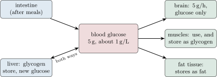
The of glucose in the blood are a pool through which large flows pass. Inputs: the intestine after a meal (up to in an hour), and between meals the liver, which releases glucose from its stored glycogen (, half a day’s supply) and makes new glucose from amino acids and lactate. Outputs: the brain (, constant), muscles (from a few grams to at work), other tissues, and storage as glycogen in the liver and muscles or as fat. A pool of fed and drained at tens of grams per hour is stable only because the flows are adjusted minute by minute.
Proof. Admitted at this level. ∎
Example 32.3 (A day of glycaemia)
Breakfast at 8: glycaemia rises from to by 9 and is back at by 10, the excess having gone into liver and muscle glycogen. From 10 to 13 the liver releases glycogen and the level holds near . A run at 17 draws into the muscles in an hour; the liver empties half its store to keep pace, and the level dips to . Overnight, the liver makes new glucose from amino acids, and at 7 the level is . Three hundred grams have passed through a pool of five.
32.2 The pancreas holds the balance
Proposition 32.4 (Two hormones from the islets)
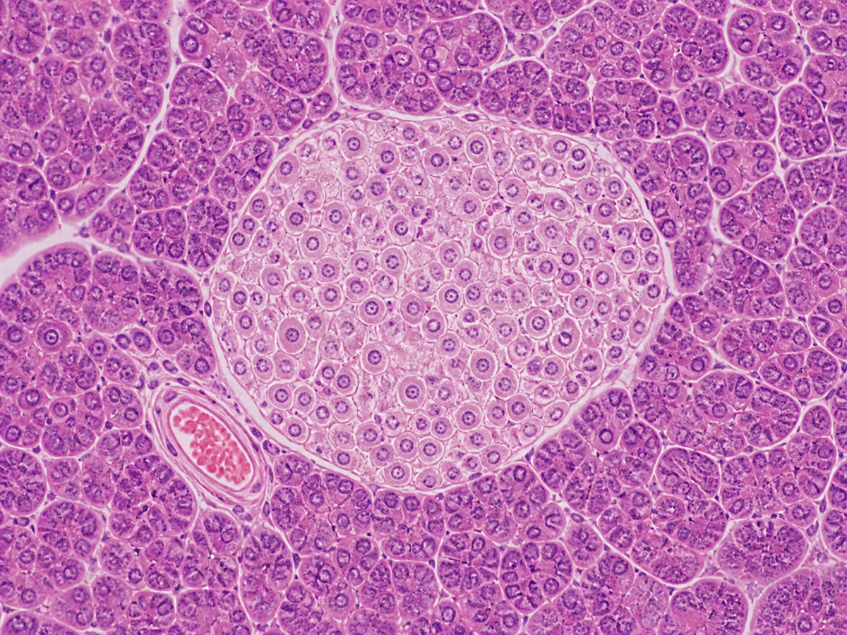
Scattered through the pancreas, among the cells that make digestive enzymes, lie a million small clusters of cells, the islets. Their cells measure the glycaemia of the blood flowing past and secrete two hormones into it:
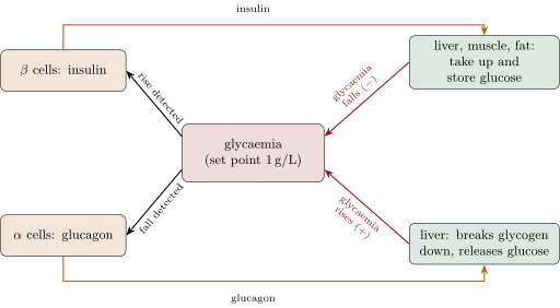
- insulin, from the cells, when the glycaemia rises: it makes muscle and fat cells take up glucose, and the liver and muscles store it as glycogen; the glycaemia falls;
- glucagon, from the cells, when it falls: it makes the liver break glycogen down and release glucose; the glycaemia rises.
The two act on the same organs in opposite directions; their balance, reset every few minutes by the measured glycaemia, is the regulation. Insulin is the only hormone that lowers the glycaemia; several raise it.
Evidence. Removing the pancreas of a dog (1889) makes it diabetic within a day: its glycaemia triples and glucose appears in its urine; tying off the duct that carries the digestive enzymes does not, so the effect is not digestive. Injecting an extract of the islets (1921) lowers the glycaemia of a diabetic dog, and of a diabetic child, within hours; the active substance, insulin, was purified the same year. Isolated islets in a dish secrete insulin when the glucose of the medium is raised and glucagon when it is lowered. Injected glucagon raises the glycaemia of a fasting person within minutes, and only if the liver holds glycogen. ∎
Example 32.5 (The loop in action)
After a meal the glycaemia climbs; within minutes the cells release insulin, the muscles and fat cells open their glucose transporters, the liver switches to making glycogen, and the level falls back — at which point insulin secretion falls too. Between meals the drift downward is met by glucagon, glycogen is broken down, and the level holds. Neither hormone is ever absent; their ratio moves, and the glycaemia follows it within a narrow band.
32.3 When the loop fails: diabetes
Definition 32.6 (Diabetes)
Diabetes is a chronic excess of glycaemia: above fasting, or above two hours after a standard glucose load. Its early signs follow from the excess: glucose in the urine, which drags water with it (abundant urine, thirst), and weight loss when cells cannot use the glucose around them. Its late damage — to the small vessels of the eye and kidney, to nerves, to the arteries — comes from years of glucose reacting with proteins. Two diseases share the name.
Proposition 32.7 (Type 1 and type 2)
- Type 1 diabetes (about 10% of cases) is the destruction of the cells by the patient’s own immune system, usually in childhood or youth: insulin is absent. The glycaemia rises unchecked, fat is burned for lack of usable glucose and its acid products accumulate; without injected insulin the disease is fatal within months. Treatment is insulin, several times a day, dosed to the meals and the measured glycaemia, for life.
- Type 2 diabetes (about 90%) develops in adults, most often overweight and inactive: the tissues respond less and less to insulin (insulin resistance), the cells compensate by secreting more, and after years they become exhausted. Insulin is present but insufficient. The predisposition is partly genetic (Chapter 16); the trigger is the way of life, and the first treatment is its change — weight loss and exercise — before drugs and, late, insulin.
Evidence. Type 1 patients have antibodies against their own islet cells years before symptoms, and at diagnosis their islets are almost empty of cells and infiltrated by white cells; their blood insulin is undetectable and they respond to injected insulin at once. Type 2 patients have normal or high insulin at diagnosis; their muscle and fat cells take up less glucose for a given insulin dose than a healthy person’s; identical twins share the disease in 70% of cases, and weight loss or a walking programme alone restores normal glycaemia in many early cases. ∎
Example 32.8 (Managing a day with type 1)
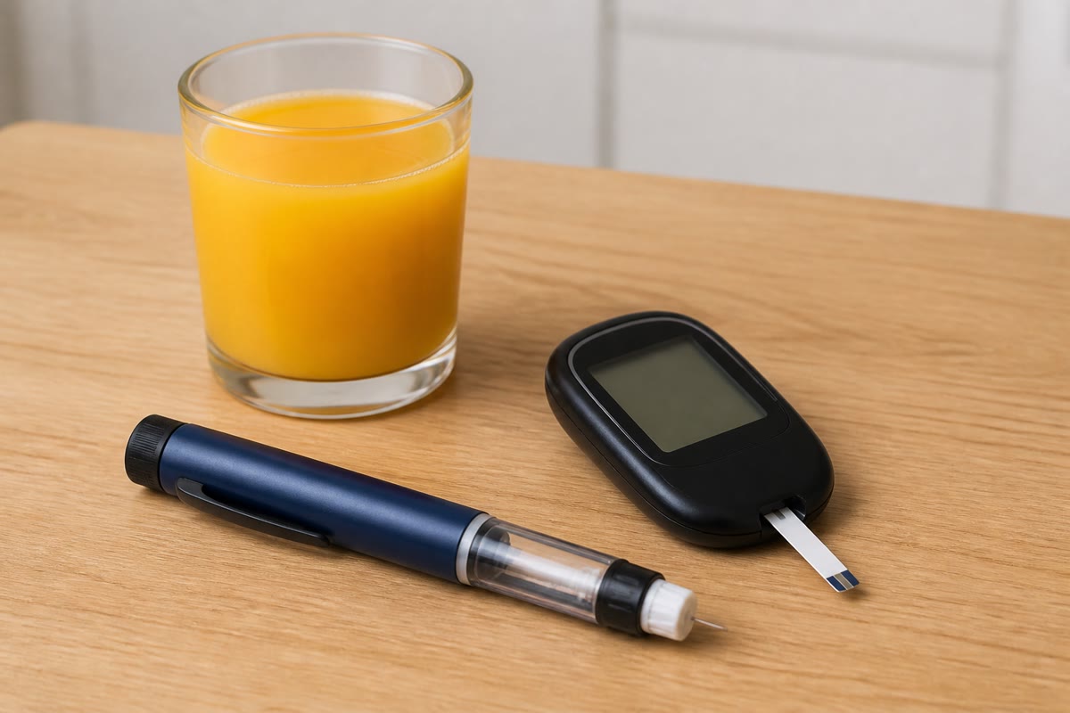
A teenager with type 1 measures her glycaemia before each meal with a drop of blood, counts the carbohydrate on her plate, and injects a dose of fast insulin proportional to it — about one unit per — plus a slow insulin once a day for the background. Too much insulin, or a missed meal, and the level falls below : sweating, trembling, confusion, corrected by sugar in minutes. Too little, and the level stays above with thirst and fatigue, and the vessels take the damage silently. She is doing, by hand, what her cells did by feedback.
Method 32.9 (Reading a glycaemia record)
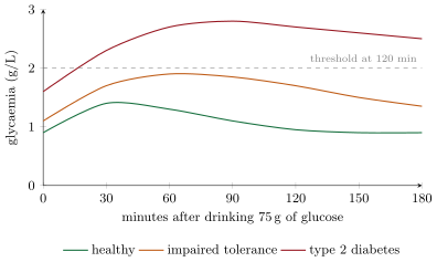
- Locate the set point and the band: fasting , after meals below ; two hours after a glucose load below (healthy), (impaired), above (diabetes).
- For each rise, ask what put glucose in (meal, liver) and what should take it out (insulin’s targets); for each fall, what took it out (brain, muscle) and what should put it back (glucagon, the liver).
- Read the hormones with the glucose: insulin should rise with the glycaemia and glucagon fall; absent insulin means type 1, high insulin with high glycaemia means resistance.
- Convert units when needed: of glucose is .
Remark 32.10 (A model of regulation)
Sensor, set point, two opposed effectors, negative feedback: the control of glycaemia is the same design as the control of testosterone in Chapter 20, and the design of most of the body’s constants — temperature, water, blood pressure, acidity. Diabetes is what a regulated constant looks like when its loop is broken: the value still responds to inputs, but nothing brings it back. Reading the curves of this chapter is practice for reading any of them.
32.4 Exercises
Exercise 32.1 ★
Give the normal fasting glycaemia in and in , and the two danger thresholds.
Solution
Solution of Exercise 32.1.
About , i.e. . Below the brain fails; above glucose passes into the urine (and years above damage the vessels).
Exercise 32.2 ★
Name the two hormones of the islets, the cells that make them, and the effect of each on the glycaemia.
Exercise 32.3 ★
Which organ can both store and release glucose? In what form does it store it?
Solution
Solution of Exercise 32.3.
The liver, as glycogen (and it can also make new glucose from amino acids and lactate).
Exercise 32.4 ★
State the essential difference between type 1 and type 2 diabetes in one sentence each.
Exercise 32.5 ★
From the tolerance-test figure, read the three curves at 120 minutes and classify each.
Solution
Solution of Exercise 32.5.
Healthy about ; impaired tolerance about (between 1.4 and 2); type 2 about (above 2).
Exercise 32.6 ★★
The brain uses of glucose per hour and the blood holds . Explain why a person does not lose consciousness an hour after a meal, naming the organ and the hormone responsible.
Exercise 32.7 ★★
Explain why removing the pancreas causes diabetes but tying its duct does not, and what this shows about where insulin is made and how it travels.
Solution
Solution of Exercise 32.7.
The duct carries digestive enzymes to the gut; blocking it leaves glycaemia normal, so the pancreas’s effect on glucose is not through digestion. Removing the whole organ removes the islets: insulin is made in the pancreas and reaches its targets through the blood, not the duct.
Exercise 32.8 ★★
A fasting person receives an injection of glucagon: the glycaemia rises by in 20 minutes. The same injection after three days of fasting does nothing. Explain.
Exercise 32.9 ★★
Why does a diabetic pass abundant urine and feel thirsty?
Solution
Solution of Exercise 32.9.
Above about the kidneys cannot reabsorb all the glucose they filter; it passes into the urine and, by osmosis, draws water with it. The water lost must be replaced: thirst.
Exercise 32.10 ★★
A type 1 patient injects her usual dose of insulin and then skips lunch. Predict what happens over the next two hours and why.
Exercise 32.11 ★★
Explain why exercise lowers the glycaemia of a type 2 diabetic even without any change in insulin, using Chapter 10.
Solution
Solution of Exercise 32.11.
A working muscle takes up glucose from the blood by a route that does not need insulin, and trained muscle regains sensitivity to insulin between sessions; the glycaemia falls during and after exercise, and the tissues’ resistance, the disease’s cause, is reduced.
Exercise 32.12 ★★★
Two patients have a fasting glycaemia of . One has no detectable insulin; the other has three times the normal insulin. Diagnose each, and say what each patient’s islets are doing.
Exercise 32.13 ★★★
Draw, or describe, the curves of glycaemia and of insulin over the three hours after a meal in a healthy person, in a type 1 patient without treatment, and in a type 2 patient. Explain each difference.
Solution
Solution of Exercise 32.13.
Healthy: glycaemia rises to about at 30–60 minutes and returns by 2 hours; insulin rises with it and falls with it. Type 1 untreated: glycaemia climbs above and stays; insulin flat at zero — nothing removes the glucose. Type 2: glycaemia rises high and falls slowly; insulin rises higher than normal and stays high — much hormone, little effect.
Exercise 32.14 ★★★
A meal contains of carbohydrate. If all of it entered the blood at once, what would the glycaemia become? Explain why it never does, listing the fates of the glucose in the first two hours.
Exercise 32.15 ★★★
Compare the regulation of glycaemia with that of testosterone in Chapter 20: sensor, set point, effectors, sign of the feedback. What is special about having two opposed hormones?
Solution
Solution of Exercise 32.15.
Both: a sensor (islet cells; hypothalamus and pituitary), a set point, effectors, and negative feedback that removes the signal. Glycaemia has two opposed hormones acting on the same organs, so the variable can be pushed both up and down actively; testosterone’s loop has one hormone and corrects mainly by more or less of it. Two opposed effectors give a faster, tighter control of a variable that changes quickly in both directions.
32.5 Problem: Five Grams of Sugar
Problem 32.1
Weekend problem — the glucose of the blood accounted for over a day: the pool and its flows, a tolerance test read, an insulin dose computed, and a loop that has to be replaced by hand
Take a blood volume of , a brain consumption of of glucose, a liver glycogen store of , and muscle glycogen of . One unit of fast insulin disposes of about of carbohydrate.
Part I — The pool.
- Compute the mass of glucose in the blood at , and the same concentration in (glucose ).
- How long would the blood’s glucose last the brain alone, if nothing replaced it?
- Between meals, the liver replaces it. How long does the liver’s glycogen last the brain? What happens after that?
- Muscle glycogen is four times the liver’s, but muscles cannot release glucose into the blood. Explain why muscle glycogen does not help the brain, and what it is for.
- A meal delivers of glucose over an hour. If none were removed, what would the glycaemia reach? What actually removes it, and where does it go?
Part II — The test. A patient drinks of glucose. Measured glycaemia and insulin:
| minutes | 0 | 30 | 60 | 90 | 120 | 180 |
|---|---|---|---|---|---|---|
| glycaemia (g/L) | 1.15 | 1.8 | 2.05 | 2.0 | 1.85 | 1.6 |
| insulin (% of a healthy peak) | 90 | 140 | 180 | 200 | 190 | 160 |
- Classify the patient with the thresholds of Method 32.9.
- Is insulin lacking? What, then, is failing?
- Name the disease, and give two features of the patient’s history you would expect.
- What would the same table look like for a type 1 patient without treatment?
- What is the first treatment prescribed, and by what mechanism does it act?
Part III — Running the loop by hand. A type 1 patient’s glycaemia is before dinner; the meal holds of carbohydrate.
- Compute the dose of fast insulin for the meal.
- She injects it but eats only half the meal. Estimate how far the glycaemia may fall, and name the symptoms and the remedy.
- She injects nothing and eats the whole meal. Estimate the glycaemia if the all entered a pool, and say what the kidneys do above .
- Why must she also inject a slow insulin at night, when she eats nothing?
- Explain in what sense the meter and the pen replace the cell, and what they cannot replace.
Part IV — The loop, reasoned.
- A drug stimulates the cells to secrete more insulin. For which type of diabetes can it work, and why not for the other?
- A person with type 2 loses and walks an hour a day; six months later the tolerance test is normal. Explain which part of the loop was repaired.
- Explain why a healthy person’s insulin and glucagon are never both high, and what an islet in a dish does when the glucose of the medium is set at .
- Glucagon, adrenaline, cortisol and growth hormone all raise the glycaemia; only insulin lowers it. Propose why the body has several safeguards against a low glycaemia and one against a high one.
- State the result: the glucose in the blood, the hours the liver can cover, the diagnosis of Part II, and the dose of Part III.
Solution
Solution of Problem 32.1.
1. ; .
2. One hour.
3. hours. After that the liver makes new glucose from amino acids (and lactate), at the cost of the body’s proteins.
4. Muscle cells lack the enzyme that releases free glucose from glycogen into the blood; their glycogen is burned on the spot, for their own contraction.
5. in : . Insulin sends the glucose into liver and muscle glycogen and into fat, and the tissues burn some; the glycaemia peaks around .
6. Fasting (below 1.26) but at 120 minutes (between 1.4 and 2): impaired tolerance, at the threshold of diabetes.
7. No: insulin is at twice a healthy peak. The tissues’ response to it is failing — insulin resistance.
8. Type 2 (or its warning stage). Expected: adult, overweight, inactive, often with diabetic relatives.
9. Glycaemia starting above , rising above and not falling; insulin near zero throughout.
10. Weight loss and regular exercise: they restore the tissues’ sensitivity to insulin and let working muscle take up glucose without it.
11. units.
12. The dose disposes of but only arrive: the surplus insulin drives the glycaemia down by the equivalent of in the pool and stores — well below : sweating, trembling, confusion; sugar at once.
13. if nothing were removed; in reality several . Above the kidneys let glucose into the urine, with water: abundant urine and thirst.
14. Between meals the liver would otherwise release glucose unchecked (glucagon acts, nothing opposes it) and the tissues would burn fat with acid products: a background of insulin is needed for the basic balance, not only for meals.
15. The meter measures the glycaemia as the cell did; the pen delivers the insulin it would have secreted. They cannot replace the continuous, minute-by-minute adjustment: the patient measures a few times a day and estimates, where the cell measured constantly and responded exactly.
16. Type 2, whose cells exist and can be pushed. In type 1 there are no cells to stimulate.
17. The tissues’ response to insulin — the effector side of the loop: with less fat and active muscle, a normal insulin signal again produces normal uptake, and the cells are relieved.
18. Each is secreted in response to the opposite deviation of the same variable: a high glycaemia calls insulin and silences glucagon, a low one the reverse. At the islet secretes both at a low, balanced rate — its resting state.
19. A low glycaemia kills the brain within minutes, a high one damages slowly over years: the acute danger has several independent guards, the slow one a single one — which is why the failure of insulin alone is enough to cause a disease, and why diabetes is common.
20. About of glucose in the blood; the liver’s glycogen covers about 20 hours; the patient of Part II has impaired tolerance at the edge of type 2 diabetes; the dose of Part III is 9 units.