High School Biology · Grades 10–12
9Muscles and Joints
A footballer plants his foot, turns, and goes down clutching his knee. The diagnosis, an hour later: a torn ligament. Nothing in the bones is broken, no muscle has failed; a strap of tissue a few centimetres long, whose job was to hold two bones in line, has given way under a twist it was never built for. To understand what tore, and why the knee will need months to recover, we need to know what a joint is made of, how muscles pull on it, and what forces run through it when a body of seventy kilograms lands on one leg.
9.1 The joint
Definition 9.1 (Joint)
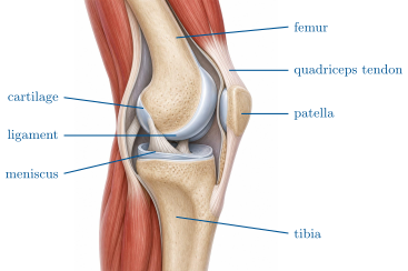
A joint is the place where two bones meet. In a mobile joint such as the knee, hip, shoulder or elbow, the ends of the bones are covered with a layer of smooth cartilage, a few millimetres thick, and enclosed in a sealed sac, the capsule, whose lining secretes the synovial fluid that lubricates the surfaces. Bands of tough tissue, the ligaments, run from bone to bone across the joint and limit its movements to the intended ones. In the knee two crescents of cartilage, the menisci, sit between the bones and spread the load.
Proposition 9.2 (What each part is for)
Cartilage and synovial fluid make the surfaces slide with less friction than ice on ice; the menisci distribute a load that would otherwise concentrate on a few square millimetres; the ligaments allow the knee to bend and straighten but resist twisting and sideways bending; the capsule keeps the fluid in. Each part has its own failure: cartilage wears (arthrosis), a meniscus tears under a twist with the knee bent, a ligament sprains or ruptures when the joint is forced beyond its range.
Proof. Admitted at this level. ∎
Example 9.3 (Sprain, tear, dislocation)
Twist an ankle on a kerb: the ligaments on the outside are stretched beyond their limit — a sprain; the joint swells as the capsule bleeds and fluid accumulates. Pull hard on a cold hamstring: some of its fibres rupture — a muscle tear. Fall on an outstretched arm: the head of the humerus may leave its socket — a dislocation, the ligaments and capsule stretched or torn as it goes. None is a broken bone; all can take longer to heal than one.
9.2 Muscles pull, in pairs
Definition 9.4 (Skeletal muscle and tendon)
A skeletal muscle is an organ made of thousands of long cells, the muscle fibres, bundled together; each fibre shortens when stimulated by a nerve. At both ends the muscle narrows into a tendon, a cord of non-contractile tissue attached to bone. A muscle can only pull, by shortening, and only along its own length: it moves a joint by pulling on the bone beyond it.
Proposition 9.5 (Antagonistic pairs)
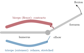
Since a muscle cannot push, every movement of a joint requires two muscles or groups working in opposition: a flexor that bends the joint and an extensor that straightens it. When one contracts, the other relaxes and is stretched; to hold a position, both contract together. The biceps flexes the elbow and the triceps extends it; the quadriceps at the front of the thigh extends the knee, the hamstrings behind it flex it.
Proof. Admitted at this level. ∎
Proposition 9.6 (Levers and forces)
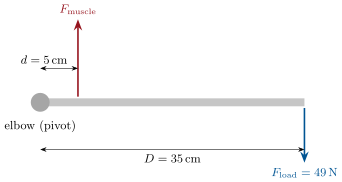
A bone pivoting at a joint is a lever. The muscle’s tendon attaches a short distance from the pivot, while the load acts at a larger distance ; for balance the muscle must pull with a force
several times the load. The arrangement trades force for speed and range: a small shortening of the muscle produces a large, fast movement of the hand or foot, at the price of large forces in the tendon and the joint.
Proof. Admitted at this level. ∎
Example 9.7 (Holding a bag)
A bag (weight ) hangs from the hand, from the elbow; the biceps tendon attaches from it. The biceps pulls with — seven times the load, the weight of — and the elbow joint is pressed together with about . Muscles, tendons and cartilage are built for these forces; what damages them is a sudden force in the wrong direction, or a load repeated beyond what the tissue can repair.
9.3 Inside the muscle
Proposition 9.8 (Structure of a muscle fibre)
A muscle fibre is a single giant cell, – wide and up to several centimetres long, with many nuclei. It is packed with parallel myofibrils, each a chain of repeating units in which two kinds of protein filament, thick and thin, interdigitate. Contraction is the sliding of the thin filaments between the thick ones, driven by ATP: every unit shortens by a fraction, the myofibril by their sum, the fibre with it. The mitochondria between the myofibrils supply the ATP; a nerve ending on each fibre gives the order.
Proof. Admitted at this level. ∎

Definition 9.9 (Fibre types)
Muscle fibres come in two main types. Slow fibres contract slowly and weakly but tirelessly: rich in mitochondria and in capillaries, red with an oxygen-storing protein, they run on respiration and dominate in postural muscles and in endurance athletes. Fast fibres contract quickly and powerfully but tire within a minute: poorer in mitochondria, paler, they rely on the phosphate store and fermentation and dominate in sprinters. The proportion in a given muscle is largely inherited; training changes the size and the equipment of the fibres more than their number.
Example 9.10 (Reading a muscle biopsy)
A stain for mitochondrial enzymes on a thin slice of thigh muscle shows a mosaic: dark fibres (slow) and pale fibres (fast). A marathon runner’s sample is 80% dark; a sprinter’s 30%; an untrained person’s about half and half. The same stain on the same runner after a year of sprint training shows the pale fibres enlarged but not more numerous.
9.4 Fatigue, damage, repair
Proposition 9.11 (Limits of the muscle)
A muscle fails in three distinct ways. Fatigue: the force declines when the ATP supply cannot keep pace — within a minute in fast fibres, over hours in slow ones — and recovers with rest. Cramp: a sudden, sustained, involuntary contraction, often from dehydration or salt loss. Injury: fibres torn by a force exceeding their strength, typically when the muscle is cold, tired, or stretched while contracting. Fibres repair over weeks from reserve cells in the muscle; a tendon or ligament, poorly supplied with blood, repairs over months.
Proof. Admitted at this level. ∎
Method 9.12 (Analysing a musculoskeletal injury)
- Identify the movement that caused it and its direction: was the joint moved within its range (a muscle problem) or beyond it (a ligament or capsule problem)?
- Identify the tissue: muscle fibres tear (pain in the belly of the muscle, weakness); ligaments sprain (pain and swelling at the joint, instability); tendons inflame under repeated load; cartilage wears silently over years.
- Estimate the force: use the lever rule; remember that landing or braking multiplies body weight several times.
- Predict the recovery from the blood supply of the tissue: muscle in weeks, ligament and tendon in months, cartilage hardly at all.
Remark 9.13 (Why warming up works)
Ten minutes of gentle movement raises the temperature of the muscles by a degree or two, which makes their proteins more extensible and their enzymes faster; it opens the capillaries that will deliver the oxygen of Chapter 8, and it thickens the synovial fluid’s film on the cartilage. A cold muscle asked for a maximal contraction is the commonest cause of a tear; a joint loaded before its fluid has spread wears faster.
9.5 Exercises
Exercise 9.1 ★
Name the parts of a mobile joint and give the role of each.
Exercise 9.2 ★
Exercise 9.3 ★
Why does every joint need at least two muscles to move?
Solution
Solution of Exercise 9.3.
A muscle can only pull by shortening; it cannot push. To move a joint both ways, a flexor and an extensor are needed, each undoing what the other did.
Exercise 9.4 ★
Distinguish a sprain, a muscle tear and a dislocation by the tissue involved.
Solution
Solution of Exercise 9.4.
Sprain: a ligament stretched or torn. Muscle tear: muscle fibres ruptured. Dislocation: a bone end forced out of its joint, with the capsule and ligaments stretched or torn.
Exercise 9.5 ★
Give two differences between slow and fast muscle fibres.
Solution
Solution of Exercise 9.5.
Slow fibres: slow, weak, tireless, rich in mitochondria and capillaries, red, run on respiration. Fast fibres: fast, powerful, tire in a minute, few mitochondria, pale, run on the phosphate store and fermentation.
Exercise 9.6 ★★
A dumbbell is held in the hand, from the elbow; the biceps attaches from it. Compute the force in the biceps ().
Solution
Solution of Exercise 9.6.
Load ; biceps force .
Exercise 9.7 ★★
Explain why the biceps and the triceps must both contract to hold the arm still against an unpredictable push.
Solution
Solution of Exercise 9.7.
A push may come in either direction, and each muscle can resist only one. Both contracting stiffen the joint in both directions at once; whichever way the push comes, one of them is already pulling against it.
Exercise 9.8 ★★
A muscle fibre long shortens by 20% when it contracts. By how much does the hand move if the fibre’s tendon attaches from the elbow and the hand is from it?
Solution
Solution of Exercise 9.8.
The fibre shortens by ; the tendon moves the bone by at from the elbow, so the hand moves .
Exercise 9.9 ★★
Why is the shoulder, the most mobile joint of the body, also the most often dislocated?
Exercise 9.10 ★★
A runner’s calf muscle cramps at the end of a hot race. Propose two causes from the chapter and one remedy.
Solution
Solution of Exercise 9.10.
Dehydration and salt loss through sweating, and fatigue of the fibres at the end of the race. Remedy: stretch the muscle gently and drink water with salt.
Exercise 9.11 ★★
Using Example 9.10, explain why a sprinter who trains for a marathon improves less than an untrained person of the same age might.
Solution
Solution of Exercise 9.11.
The sprinter’s thigh is 70% fast fibres and the proportion is largely inherited; training enlarges fibres and improves their equipment but does not convert their number. The untrained person starts from half and half and has more slow fibres to develop.
Exercise 9.12 ★★★
When landing from a jump the knee bends and the quadriceps tendon carries about four times the body weight. For a athlete, compute that force. The tendon has a cross-section of about and fails at about ; how far is it from failure?
Solution
Solution of Exercise 9.12.
. Stress : about a quarter of the failure stress, a safety factor of four.
Exercise 9.13 ★★★
A torn ligament heals over months, a torn muscle over weeks, and damaged cartilage barely at all. Relate the three to the blood supply of the tissues and to the cells available for repair.
Solution
Solution of Exercise 9.13.
Muscle is richly supplied with blood and contains reserve cells that rebuild fibres: weeks. Ligaments have few vessels, so nutrients and repair cells arrive slowly: months. Cartilage has no blood vessels at all and almost no dividing cells; it is fed by the synovial fluid only and barely repairs.
Exercise 9.14 ★★★
Bodybuilding enlarges the muscles; stretching lengthens the range of a joint. Which tissue does each one change, and why does neither change the number of fibres?
Solution
Solution of Exercise 9.14.
Bodybuilding thickens the fibres (more myofibrils in each) and stretching lengthens the muscle–tendon unit and the capsule’s tolerance. Fibre number is fixed early in life; adult muscle grows or shrinks by changing the size of existing fibres.
Exercise 9.15 ★★★
An elderly person’s knee cartilage has worn to half its thickness. Explain, from the chapter, why the joint hurts on stairs, why the menisci matter more than before, and why the muscles around the knee are part of the treatment.
Solution
Solution of Exercise 9.15.
On stairs the joint carries several times body weight; thinner cartilage transmits that pressure to the bone beneath, which has nerves and hurts. With less cartilage the menisci are the last thing spreading the load. Strong muscles around the knee absorb part of the shock and steady the joint, reducing the peak pressure on what cartilage remains.
9.6 Problem: The Forces in a Knee
Problem 9.1
Weekend problem — a footballer’s knee taken apart: the lever of the leg, the pull of the thigh muscle, the ligament that tore, and the numbers a tendon carries on every step
Karim, , injured his knee turning sharply on a planted foot. Take . The quadriceps tendon attaches to the tibia from the knee’s pivot; the ground’s push on the foot acts about from it when the knee is well bent, as in a landing.
Part I — Standing and stepping.
- Compute Karim’s weight.
- Standing on one leg with the knee slightly bent, the ground pushes up on the foot with his whole weight, acting from the knee’s pivot. Compute the force in the quadriceps tendon.
- Climbing a stair, the knee is bent further and the ground force acts from the pivot. Compute the tendon force again.
- Express the two results as multiples of body weight, and explain why stairs tire the thighs.
- Which muscle group is the antagonist of the quadriceps, and what is it doing during the step?
Part II — The landing.
- Landing from a header, the ground pushes with three times Karim’s weight, from the pivot. Compute the tendon force.
- The tendon’s cross-section is . Compute the stress in it, in newtons per square millimetre, and compare with the failure stress of about .
- The joint surfaces are pressed together by about the tendon force plus the ground force. Compute that load, then the pressure on the cartilage if the contact area is with the menisci and without them.
- Deduce why a knee that has lost a meniscus wears its cartilage faster.
- Explain what the synovial fluid contributes during the landing.
Part III — The injury.
- Karim’s foot was planted and his body turned: which direction of movement was imposed on the knee, and which tissue is built to resist it?
- Why did the quadriceps, strong enough to pull with several kilonewtons, not protect the ligament?
- The knee swelled within an hour. What filled it, and which part of the joint was breached?
- A scan shows the ligament ruptured and a meniscus torn. Using Method 9.12, predict the recovery time of each and justify from their blood supply.
- Karim asks why the surgeon wants to strengthen his hamstrings before the operation. Answer with Proposition 9.5.
Part IV — Every step counts.
- Karim takes about 8000 steps a day; half are on the injured leg. Using question 2’s force, how many times per day does the tendon carry at least that load?
- Over a year, what is the total number of loadings? Explain why a tendon inflamed by overuse (tendinitis) needs rest rather than strength.
- During a match he lands from jumps some 40 times. Compare the total load carried in landings (question 6) with that carried in the day’s steps.
- Running doubles the ground force compared with walking. Why do doctors recommend cycling and swimming rather than running for a healing knee?
- State the result: the multiple of body weight carried by the quadriceps tendon on a stair and on a landing, and the one tissue of the knee whose strength those numbers never tested — until the turn.
Solution
Solution of Problem 9.1.
1. .
2. .
3. .
4. Two and four times body weight: on every stair the quadriceps pulls with the weight of four people, which is why thighs burn on a long climb.
5. The hamstrings, at the back of the thigh; they relax and are stretched while the quadriceps straightens the knee, contracting lightly to steady the joint.
6. Ground force ; tendon force , twelve times body weight.
7. : about two thirds of the failure stress. Each landing uses most of the tendon’s margin.
8. Load . With the menisci: ; without: , six times more.
9. Six times the pressure on the same cartilage at every landing: the surface is crushed faster than it can be maintained, and it wears.
10. A film of fluid between the cartilages, so that they slide during the bend without dry friction, and a slight cushioning of the impact.
11. A twist of the tibia under the femur, with a sideways bend: rotations the knee is not built for, which the ligaments are there to resist.
12. The quadriceps pulls only along the leg, straightening the knee; it has no action against a twist. And the injury happens in a few hundredths of a second, faster than any muscular response.
13. Blood from the torn ligament’s and capsule’s vessels, plus extra synovial fluid: the capsule was breached or inflamed and the joint filled.
14. The ligament, poorly supplied with blood, needs months and often surgery; the torn meniscus, cartilage almost without vessels, usually does not heal and the torn part is trimmed or sutured.
15. The hamstrings are the quadriceps’ antagonists and pull the tibia backwards; a strong pair contracting together stiffens the knee against the forward slide the torn ligament no longer prevents, protecting the joint before and after surgery.
16. About 4000 times a day at or more.
17. About loadings a year. Tendinitis is damage accumulating faster than the slow repair of tendon tissue; only rest lets repair catch up, since strength does not reduce the load of each step.
18. Landings in all; steps — the steps carry seventeen times more total load, but the landings carry the peaks that threaten rupture.
19. Doubling the ground force doubles the tendon and joint forces at every stride; cycling and swimming work the muscles while loading the knee far below body weight.
20. The quadriceps tendon carries four times body weight on a stair and about twelve on a landing; the ligaments, which those loads never test, were the tissue the twist found.