High School Biology · Grades 10–12
35The Stretch Reflex and the Nerve Message
A doctor taps the tendon just below your kneecap with a small rubber hammer, and your foot kicks forward before you have felt anything. Thirty milliseconds separate the tap from the kick: too little for the brain, too little even for a decision. The signal went from the muscle to the spinal cord and back — a circuit of two neurons and one connection between them — and the same circuit is what holds you upright, silently, every second you stand. This chapter uses it to introduce the neuron, the electrical signal it carries, and the chemical junction where one neuron speaks to the next.
35.1 The reflex arc
Definition 35.1 (Reflex)
A reflex is an involuntary, rapid and stereotyped response of an effector (a muscle or a gland) to a stimulus, produced by a fixed circuit of neurons, the reflex arc, that runs through the spinal cord or brainstem without requiring the brain. The stretch reflex is the simplest: the sudden stretching of a muscle makes that same muscle contract.
Proposition 35.2 (The circuit of the stretch reflex)
Five elements, in order:
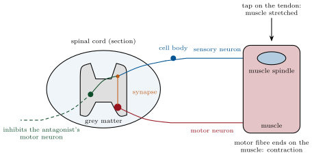
- the receptor: muscle spindles, small sensory organs lying among the muscle fibres, which are stretched when the muscle is and respond by firing signals;
- the sensory neuron, whose fibre runs from the spindle to the spinal cord, entering by the dorsal root; its cell body sits in a ganglion beside the cord;
- the integration centre: the grey matter of the spinal cord, where the sensory fibre connects directly — through a single synapse — to the motor neuron of the same muscle, and, through an intermediate neuron, inhibits the motor neurons of the antagonist muscle;
- the motor neuron, whose fibre leaves by the ventral root and runs to the muscle;
- the effector: the muscle fibres, which contract.
A tap on the patellar tendon stretches the thigh muscle; its spindles fire; the motor neurons of that muscle fire; it contracts and the leg extends, while the hamstrings are told to relax.
Evidence. Cutting the dorsal root abolishes the reflex while the muscle can still be made to contract by stimulating the ventral root; cutting the ventral root abolishes it while the sensory fibres still carry signals when the muscle is stretched: the arc has a sensory entrance and a motor exit. The reflex persists in an animal whose spinal cord has been severed from the brain: the brain is not needed. Recording from single fibres shows the spindle’s signals starting within a millisecond of the stretch, and the motor neuron firing about one millisecond after the sensory signal arrives — the delay of one synapse. ∎

Example 35.3 (What the reflex is for)
Standing, you sway; each sway stretches the muscles of one side of the ankle, whose spindles fire and whose reflex contraction pulls you back before you notice. Carrying a tray, a sudden extra weight stretches the arm muscles and the reflex stiffens them within , before the tray tips. The stretch reflex is a servomechanism: it opposes any change of a muscle’s length, and the brain adjusts its sensitivity to set the body’s tone — higher when you brace, lower when you sleep.
35.2 The neuron and its message
Definition 35.4 (Neuron)
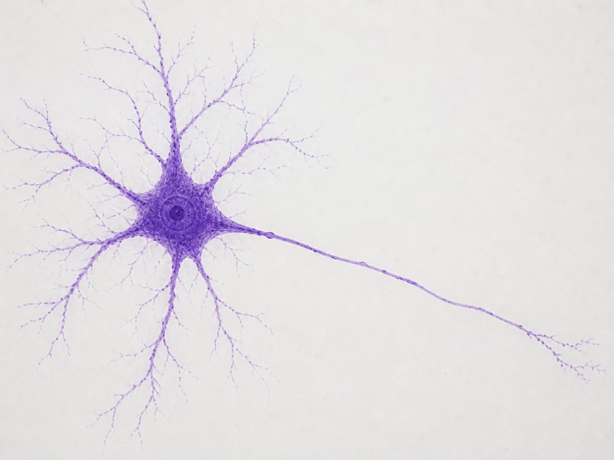
A neuron is a cell specialised for signalling: a cell body with its nucleus, branching dendrites that receive signals, and a single long fibre, the axon, that carries the neuron’s own signal to its endings — for a motor neuron of the leg, a metre away. Many axons are wrapped in a fatty sheath, myelin, laid down by companion cells, which speeds conduction. A nerve is a bundle of hundreds or thousands of axons.
Proposition 35.5 (The action potential)
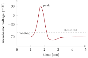
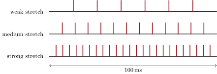
A neuron’s membrane holds an electrical voltage: the inside is about relative to the outside at rest. The nerve message is a train of action potentials: brief reversals of that voltage, each rising to about and returning within a millisecond, that travel along the axon without weakening. An action potential is all or none: a stimulus below a threshold produces nothing, any stimulus above it produces the same full-sized signal. The intensity of a stimulus is therefore coded not in the size of the signals but in their frequency: a stronger stretch makes the spindle fire more action potentials per second.
Evidence. Microelectrodes inserted into an axon record the resting voltage and, when the cell fires, identical spikes whose shape does not change with the stimulus or with the distance travelled. Recordings from a single spindle fibre during increasing stretches show spikes of constant size at rates rising from a few to several hundred per second. A stimulus just below threshold gives no spike at all; doubling a stimulus already above threshold changes the rate, never the height. ∎
Example 35.6 (Speed)
Action potentials travel at in thin unmyelinated fibres and up to in the thick myelinated fibres of the stretch reflex. From the knee to the spinal cord and back is about : some of travel, to which the spindle’s response, one synapse and the muscle’s activation add another — the of the knee jerk. A message from a toe to the brain, away, takes about ; the decision to move it, and its execution, some more.
35.3 The synapse
Definition 35.7 (Synapse and neurotransmitter)
A synapse is the junction where the ending of one neuron’s axon meets the next cell — a neuron or a muscle fibre — across a gap of some , the synaptic cleft. The signal does not jump the gap electrically: the arriving action potentials make the ending release, from small vesicles, a chemical messenger, the neurotransmitter, into the cleft; it binds receptors on the receiving cell’s membrane, which change that cell’s voltage; it is then destroyed or taken back within milliseconds. The synapse converts an electrical message into a chemical one and back, and the amount of transmitter released — set by the frequency of the arriving action potentials — codes the message’s intensity.
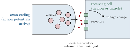
Proposition 35.8 (Excitation, inhibition, and the muscle’s junction)
Depending on the transmitter and its receptor, a synapse excites the receiving neuron (brings it towards its threshold) or inhibits it (drives it away). The stretch reflex uses both: excitation of the muscle’s own motor neuron, inhibition of the antagonist’s through the interneuron. At the neuromuscular junction, the synapse between a motor neuron and a muscle fibre, the transmitter is acetylcholine: each action potential of the motor neuron releases enough to fire the fibre, which contracts. A motor neuron and the fibres it commands form a motor unit; the force of a muscle is set by how many units fire and how fast.
Evidence. Acetylcholine applied to a muscle fibre makes it contract; curare, the arrow poison, binds the fibre’s acetylcholine receptors without activating them and paralyses every muscle while the nerves keep firing; the toxin of botulism blocks the release of the vesicles and paralyses likewise; an insecticide that blocks the enzyme destroying acetylcholine makes the muscles contract uncontrollably. Strychnine blocks the inhibitory synapses of the spinal cord, so that every stretch triggers the contraction of both muscle and antagonist: convulsions. Each poison names a step of the junction by removing it. ∎
Method 35.9 (Analysing a reflex or a poison)
- Trace the arc: receptor, sensory neuron, centre, motor neuron, effector; identify which element a lesion or a drug affects.
- At each neuron, ask what is coded — the frequency of action potentials — and at each synapse, what carries the message — the transmitter and its amount.
- For a poison, find its step: release of the transmitter, the receptor, the destruction of the transmitter, or an inhibitory synapse; predict paralysis (nothing gets through) or convulsion (nothing is stopped).
- Time it: distance divided by conduction speed, plus about a millisecond per synapse.
Remark 35.10 (A message made of the same spikes)
Every message in the nervous system — the spindle’s stretch, the retina’s light, the brain’s command to move — is made of the same all-or-none action potentials, differing only in rate and in the wires that carry them; meaning lies in which neuron fires, not in what it fires. And at every synapse the message becomes a dose of a chemical, which is why a molecule — a transmitter, a poison, a drug — can enter the conversation at any point. The final chapter follows the same spikes and synapses up into the brain, and the command that comes back down.
35.4 Exercises
Exercise 35.1 ★
List the five elements of the stretch reflex arc, in order.
Exercise 35.2 ★
Describe a neuron’s parts and the direction in which its message travels.
Exercise 35.3 ★
What is an action potential? What does "all or none" mean, and how is intensity coded?
Solution
Solution of Exercise 35.3.
A brief reversal of the membrane voltage, from to about and back within a millisecond, travelling along the axon unchanged. All or none: below threshold nothing, above it always the same full signal. Intensity is coded in the frequency of action potentials.
Exercise 35.4 ★
Describe the events at a synapse from the arrival of action potentials to the response of the receiving cell.
Solution
Solution of Exercise 35.4.
Action potentials reach the ending; vesicles release the neurotransmitter into the cleft; it binds receptors on the receiving cell, changing its voltage (towards or away from threshold); it is then destroyed or taken back.
Exercise 35.5 ★
Which transmitter acts at the neuromuscular junction, and what do curare and botulinum toxin each do to it?
Solution
Solution of Exercise 35.5.
Acetylcholine. Curare occupies its receptors on the muscle without activating them; botulinum toxin prevents its release from the vesicles. Both paralyse.
Exercise 35.6 ★★
From the frequency-coding figure, count the spikes in for each stretch and give the rates. What does not change between the rows?
Solution
Solution of Exercise 35.6.
6, 12 and 24 spikes in : 60, 120 and 240 per second. The size and shape of the spikes do not change.
Exercise 35.7 ★★
The knee-jerk latency is for a path of . If the synapse and the muscle’s activation take together, compute the conduction speed of the fibres.
Solution
Solution of Exercise 35.7.
Conduction time for : about .
Exercise 35.8 ★★
Explain why cutting the dorsal root abolishes the reflex but not the muscle’s ability to contract, and cutting the ventral root abolishes both.
Solution
Solution of Exercise 35.8.
The dorsal root carries the sensory fibres: cut, the stretch message never reaches the cord, but the motor neuron and its fibre are intact and can still be stimulated to contract the muscle. The ventral root carries the motor fibres: cut, nothing can reach the muscle from the cord at all.
Exercise 35.9 ★★
Why must the antagonist muscle be inhibited during the reflex? Which element of the arc does it, and through what kind of synapse?
Exercise 35.10 ★★
A stimulus of 2 units is just at a neuron’s threshold and gives one action potential of . Predict the signal for stimuli of 1, 4 and 8 units.
Solution
Solution of Exercise 35.10.
1 unit: nothing (below threshold). 4 and 8 units: action potentials of the same , but at higher frequencies, higher for 8 than for 4.
Exercise 35.11 ★★
Explain why the message crosses a synapse in one direction only, and why it is delayed there by about a millisecond.
Solution
Solution of Exercise 35.11.
Only the ending holds vesicles of transmitter and only the receiving membrane holds receptors, so the chemical can act one way. Releasing the transmitter, its diffusion across the cleft and the receptors’ response take about a millisecond.
Exercise 35.12 ★★★
An insecticide blocks the enzyme that destroys acetylcholine. Predict its effect at the neuromuscular junction, on breathing, and explain why a drug that blocks the acetylcholine receptors is used as an antidote at a carefully measured dose.
Solution
Solution of Exercise 35.12.
Acetylcholine accumulates in the junctions and keeps firing the fibres: twitching, then sustained contraction and exhaustion of the muscles, including those of breathing — death by asphyxia. A receptor blocker (an atropine-like drug) reduces the excess stimulation; too little leaves the poisoning, too much paralyses like curare, so the dose must match.
Exercise 35.13 ★★★
Strychnine blocks inhibitory synapses in the spinal cord. Using the reflex arc, explain why a light touch then triggers convulsions of the whole body.
Solution
Solution of Exercise 35.13.
Every sensory input excites its motor neurons and, normally, inhibits the antagonists’ through interneurons. With inhibition blocked, a touch excites both sides of every joint and the excitation spreads unopposed through the cord: all muscles contract at once, and the spasm stretches other muscles whose reflexes add to it.
Exercise 35.14 ★★★
A person with damaged myelin has a knee-jerk latency of instead of 30. Compute the conduction speed implied (using the figures of exercise 7) and explain why myelin matters.
Solution
Solution of Exercise 35.14.
Conduction time for : about , a third of normal. Myelin lets the action potential jump between gaps in the sheath instead of travelling continuously; without it the fibre conducts like a thin unmyelinated one.
Exercise 35.15 ★★★
"The spinal cord is only a cable between brain and body." Rewrite correctly in a paragraph, using the reflex, the interneuron, and the animal whose cord is cut from its brain.
Solution
Solution of Exercise 35.15.
The cord carries messages between brain and body, but it also processes them: its grey matter holds the synapses of the reflex arcs, where a stretch is turned into a contraction and, through interneurons, into the inhibition of the antagonist, without any message reaching the brain. An animal whose cord is cut from its brain still withdraws a paw from a pinch and still shows the knee jerk: the cord is a centre, not only a cable.
35.5 Problem: Thirty Milliseconds
Problem 35.1
Weekend problem — the knee jerk timed and taken apart: the spindle’s code, the fibres’ speed, the synapse’s delay, the junction’s chemistry, and the poisons that name each step
Electrodes record the electrical activity of a subject’s thigh muscle (an electromyogram) while the patellar tendon is tapped. The muscle’s signal begins after the tap; the leg moves later. The distance from the tendon to the spinal cord along the nerve is , the same back.
Part I — The timing.
- Compute the total length of the nerve path of the reflex.
- Take the spindle’s response and the muscle’s activation to need each, and the synapse . How much of the is conduction, and what is the conduction speed?
- In a subject taller, with the same fibres, predict the latency.
- Why does the leg move after the muscle’s electrical signal begins?
- A subject is told to expect the tap and to "think about" stopping the kick; the kick is unchanged. Explain with the timing of a message to and from the brain ( more of fibres, and several synapses).
Part II — The code. The spindle fibre fires 20 action potentials per second at rest, 200 during the tap.
- How many action potentials does the spindle send in the following the tap? What changes between rest and tap, and what does not?
- Each action potential arriving at the synapse releases a fixed quantity of transmitter. By what factor does the transmitter released per second rise during the tap?
- The motor neuron fires only if enough transmitter arrives within a few milliseconds. Explain why the resting rate does not make the muscle contract, and the tap rate does.
- A gentler tap gives 100 per second. Predict the motor neuron’s response and the strength of the kick, using motor units.
- Explain why a spindle’s message could not be coded in the size of its action potentials.
Part III — The junction.
- Name the transmitter at the neuromuscular junction and describe its fate after acting.
- Curare is injected: the electromyogram shows no signal after the tap, but the nerve still carries action potentials. Which step is blocked?
- Botulinum toxin, instead: same electromyogram, same nerve signal. Which step, and how would you tell the two apart in the laboratory?
- An anaesthetic blocks action potentials in the sensory fibres. Which recordings disappear, and which remain?
- Explain why all three produce a limp leg but by three different mechanisms.
Part IV — The centre.
- During the kick the hamstrings’ electromyogram is silent, and drops below its resting level. Explain with the interneuron.
- A patient with a spinal cord severed above the level of the reflex still has a knee jerk — exaggerated. What does the presence of the reflex show, and what does its exaggeration suggest about the brain’s normal role?
- In a patient whose reflex is absent on one side, list the elements of the arc that could be damaged, and one test to locate the damage.
- Why do doctors test this reflex routinely, in a few seconds, on every patient?
- State the result: the conduction speed of the reflex’s fibres, the quantity that codes the strength of the stretch, and the one chemical step where curare acts.
Solution
Solution of Problem 35.1.
1. .
2. of conduction: .
3. About more each way, in all: more, some .
4. The electrical signal is the fibres’ activation; the contraction itself — the sliding of filaments and the pull on the tendon — takes some tens of milliseconds to develop enough force to move the leg.
5. A message from the cord to the brain and a command back travel at about — — plus several synapses and the brain’s own processing: at least , and a decision far longer. The kick is over before any brain signal can reach the motor neurons.
6. At 200 per second, 2 action potentials in (0.2 at rest). The rate changes; the size and shape of each spike do not.
7. By a factor of 10.
8. At 20 per second the doses of transmitter are apart and each fades before the next; the motor neuron never reaches threshold. At 200 per second they arrive apart and add up: threshold is crossed and the motor neuron fires.
9. Fewer motor neurons reach threshold and each fires fewer action potentials: fewer motor units contract, at a lower rate, and the kick is weaker.
10. Action potentials are all or none: their size is fixed by the neuron, so it carries no information about the stimulus. Only the rate can vary.
11. Acetylcholine; after binding the receptors it is destroyed within milliseconds by an enzyme in the cleft.
12. The receptors of the muscle fibre: acetylcholine is released but cannot act.
13. The release of acetylcholine from the vesicles. In the laboratory, acetylcholine applied directly to the muscle would make it contract under botulinum toxin (receptors free) but not under curare (receptors blocked).
14. The sensory fibre’s action potentials and, since nothing reaches the cord, the motor neuron’s and the electromyogram; the muscle still contracts if its nerve is stimulated below the block.
15. Curare blocks reception, botulinum toxin blocks release, the anaesthetic blocks conduction: three steps, one silent muscle.
16. The sensory fibre also excites an interneuron that inhibits the hamstrings’ motor neurons; their resting activity is suppressed, and the antagonist relaxes below its resting tone.
17. The reflex needs only the cord, which is intact at that level. Its exaggeration shows that the brain normally sends signals that damp the reflex; without them the arc runs at full sensitivity.
18. The spindle, the sensory nerve or dorsal root, the cord’s grey matter at that level, the motor nerve or ventral root, the muscle itself. Stimulating the motor nerve directly and recording the muscle tells whether the motor side works; recording the sensory nerve during a stretch tells whether the sensory side does.
19. It tests, in one tap, the whole arc — sensory nerve, cord, motor nerve, muscle — and the brain’s damping of it: an absent, weak or exaggerated reflex points to a level of the nervous system before any other sign.
20. About ; the frequency of the spindle’s action potentials; the binding of acetylcholine to the muscle’s receptors.