High School Biology · Grades 10–12
36From Intention to Movement
A woman wakes one morning unable to move her right arm or leg, though she can feel them, and her right knee jerks harder than ever when the doctor taps it. The scan shows a small dead patch in the left half of her brain, near the top. Everything below is intact — the nerves, the spinal cord, the reflex arcs of the last chapter, the muscles — and yet the intention to move does not reach them. Six months of rehabilitation later, she walks, and the scan of her working brain shows regions lighting up that did not command her leg before. This chapter follows a voluntary movement from the cortex to the motor neuron, and shows how the map that commands it is drawn, crossed, and redrawn.
36.1 The motor cortex
Definition 36.1 (Motor cortex)
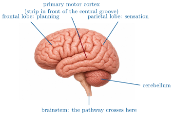
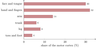
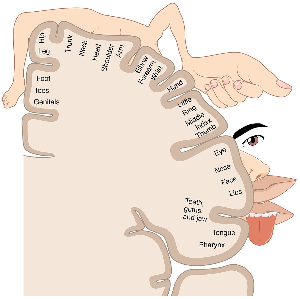
The primary motor cortex is a strip of the cerebral cortex running over the top of each hemisphere, just in front of the groove that separates the frontal lobe from the parietal. Its neurons are the origin of voluntary movement: their axons descend through the brainstem and the spinal cord to the motor neurons of the muscles. The strip is a map of the body: each part of it commands one part of the body, in an order that runs from the foot at the top of the hemisphere to the face at its side, and each part’s area is proportional not to the size of the body part but to the fineness of its movements — the hand and the face take up most of it.
Proposition 36.2 (The map is real, and crossed)
Stimulating a point of the motor cortex electrically produces a movement of the corresponding body part on the opposite side of the body; damage to a point abolishes voluntary movement of that part on the opposite side.
Evidence. During brain surgery under local anaesthesia, Penfield (1930s) applied a weak current to points of the exposed cortex and recorded which muscles moved: the points formed the map, with the hand and face areas out of proportion to their size, and every movement on the side opposite the stimulated hemisphere. Functional imaging of healthy volunteers moving one finger shows a spot of activity at the predicted place in the opposite hemisphere; moving the foot, a spot at the top. Strokes destroying part of one motor cortex paralyse the mapped parts on the other side, with sensation intact. ∎
36.2 The descending pathway
Proposition 36.3 (From the cortex to the motor neuron)
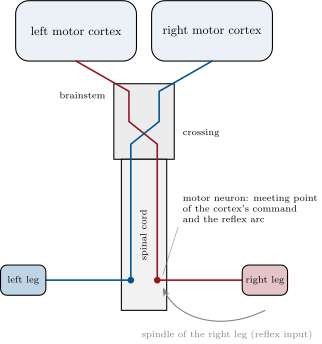
The axons of the motor cortex neurons form the corticospinal tract. It descends through the brainstem, where most of its fibres cross to the other side — which is why each hemisphere commands the opposite half of the body — and continues down the spinal cord, where at each level fibres leave to end on the motor neurons of that level’s muscles, or on interneurons beside them. The motor neuron of Chapter 35 is thus the meeting point of two commands: the reflex arc’s, from the muscle spindle, and the cortex’s, from a metre above.
Evidence. Tracing the fibres from the cortex through the brainstem shows the crossing; a lesion of the tract above the crossing paralyses the opposite side, below it the same side. A lesion of the spinal cord paralyses everything below its level on both sides while sparing the face, whose fibres leave above; the reflexes below the lesion persist, since their arcs are intact, and become exaggerated, since the cortex’s damping influence no longer reaches them. ∎
Example 36.4 (Reading a paralysis)
Right arm and leg paralysed, face too, sensation kept: a lesion of the left hemisphere’s motor cortex or its fibres above the crossing — the woman of the opening. Both legs paralysed, arms and face spared, reflexes of the legs exaggerated: the spinal cord damaged at the level of the lower back. One arm paralysed and limp, its reflexes absent: the nerve or the motor neurons themselves, below which nothing can fire. The map, the crossing and the arc locate the damage before any scan.
36.3 The motor neuron decides
Proposition 36.5 (Integration at the motor neuron)
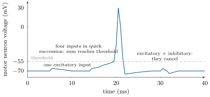
A motor neuron receives thousands of synapses: excitatory ones from the cortex and from the spindles, inhibitory ones from interneurons driven by the antagonist’s spindles, by the cortex, and by other centres. Each arriving signal shifts the neuron’s voltage a little, towards threshold or away from it; the shifts add up over milliseconds and over the cell’s surface, and the neuron fires only when their sum crosses threshold. The motor neuron is therefore not a relay but a calculator: the muscle contracts when the balance of all the commands reaching its motor neurons says so — the final common pathway of movement.
Evidence. Recording inside a motor neuron shows each excitatory input producing a small depolarisation of a millivolt or so, far below the needed; a burst of inputs, or inputs from several sources at once, summed to threshold produce an action potential; an inhibitory input arriving at the same moment cancels an excitatory one. The strength of a voluntary contraction rises with the number of motor neurons recruited and with their firing rate, and the knee jerk is stronger when the subject clenches a fist — the cortex’s excitation adding to the spindle’s. ∎
Example 36.6 (Holding a glass)
Lifting a full glass, the cortex sends a steady excitation to the motor neurons of the arm; the spindles, stretched as the glass tips, add theirs; the antagonists’ interneurons subtract when the movement overshoots; the cerebellum, comparing the intended and the actual movement, corrects the cortex’s command a few tens of milliseconds later. Each motor neuron sums the whole conversation and fires just enough. The smoothness of the movement is the balance of the sums.
36.4 A map that can be redrawn
Proposition 36.7 (Plasticity of the motor cortex)
The motor map is not fixed. Practising a movement enlarges the cortical area that commands it and sharpens its connections; a limb’s area shrinks when the limb is immobilised, and after an amputation is taken over by the neighbouring parts. After a stroke, undamaged cortex around the lesion, and the corresponding area of the other hemisphere, can learn to command the affected muscles — provided they are used: rehabilitation works by forcing the movement, repeatedly, so that the surviving circuits are selected and strengthened. The plasticity of Chapter 22 applies to the commanding side of the brain as to the sensing side.
Evidence. Imaging the cortex of violinists shows an enlarged area for the fingers of the left hand, in proportion to the age at which they began; after five days of practising a finger sequence, ordinary volunteers show a measurably enlarged area for those fingers, which shrinks again if practice stops. After a stroke, patients whose paralysed arm is trained intensively — the good arm restrained — recover more function than those who compensate with the good arm, and imaging shows the recovered movement commanded by regions neighbouring the lesion. Monkeys whose finger area is lesioned recover finger movement only if the fingers are exercised. ∎

Example 36.8 (Six months after the stroke)
Week 1: the right leg does not move; its reflexes are exaggerated. Weeks 2–6: the physiotherapist moves the leg, then makes the patient attempt each movement; a faint contraction returns. Months 2–4: walking between bars, a thousand steps a day; imaging shows activity spreading in the cortex around the lesion and in the other hemisphere. Month 6: walking with a stick. The lesion has not healed — neurons of the cortex do not regrow — but neighbouring circuits have been recruited, and each step of the recovery was made by attempting the movement.
Method 36.9 (Locating a motor disorder)
- Which parts are affected? One side of the body, face included: the opposite hemisphere or its fibres above the crossing. Both sides below a level, face spared: the spinal cord at that level. One limb or muscle group only: its nerve or motor neurons.
- Are the reflexes preserved? Exaggerated: the arc is intact and the cortical damping is lost (a lesion above the motor neuron). Absent: the arc itself is broken (the motor neuron, nerve or muscle).
- Is sensation preserved? A pure motor deficit points to the motor pathway; a mixed one to a lesion affecting both.
- What can be recovered? Cortical lesions, through plasticity and training; a severed cord or nerve, much less; muscle and motor neuron diseases, according to their cause.
Remark 36.10 (The year’s last loop)
The intention to move is a pattern of action potentials in a strip of cortex; it descends a metre of axon, crosses, and is weighed by a motor neuron against the reflex of the muscle it will move; the muscle contracts by the sliding of filaments, paid for by the ATP of the mitochondria, on glucose the liver released under the eye of the pancreas, on oxygen the heart and lungs delivered — and the cortex that gave the order was shaped, by its own use, into the map it is. Every chapter of this book has been a piece of that loop. The volumes that follow open each piece further; none of them changes the loop.
36.5 Exercises
Exercise 36.1 ★
Where is the primary motor cortex, and what does its map represent?
Solution
Solution of Exercise 36.1.
A strip over the top of each hemisphere, in front of the groove between frontal and parietal lobes. Its map assigns each part of the strip to one part of the opposite side of the body, with area proportional to the fineness of that part’s movements.
Exercise 36.2 ★
Describe the corticospinal tract from the cortex to a leg muscle, naming where it crosses.
Solution
Solution of Exercise 36.2.
Axons of motor cortex neurons descend through the brainstem, where most cross to the other side, then down the spinal cord to the level of the leg, where they end on the motor neurons (or neighbouring interneurons) whose axons run to the leg muscle.
Exercise 36.3 ★
Why does the hand take up more of the motor cortex than the whole trunk?
Solution
Solution of Exercise 36.3.
Area follows the precision of control, not size: the hand makes many fine, independent movements that need many cortical neurons; the trunk makes few and coarse ones.
Exercise 36.4 ★
What does "final common pathway" mean for the motor neuron?
Solution
Solution of Exercise 36.4.
Every command to a muscle — from the cortex, the reflex arcs, the interneurons — must pass through its motor neurons, which sum all the inputs and fire only if the sum reaches threshold; there is no other route to the muscle.
Exercise 36.5 ★
Give two pieces of evidence that the motor map can change.
Solution
Solution of Exercise 36.5.
The enlarged finger area of musicians (and of volunteers after days of practice); the takeover of an amputated limb’s area by neighbours; the recovery after a stroke commanded by regions around the lesion.
Exercise 36.6 ★★
From the integration figure, how much does one excitatory input depolarise the neuron, how many are needed within a few milliseconds to reach threshold, and what happens when an inhibitory input coincides with an excitatory one?
Solution
Solution of Exercise 36.6.
About ; four inputs in quick succession reach the threshold; an inhibitory input arriving with an excitatory one cancels it and the voltage barely moves.
Exercise 36.7 ★★
A patient cannot move the left side of the face and the left arm; the left leg is weak. Locate the lesion, using the map and the crossing.
Solution
Solution of Exercise 36.7.
Face and arm on the left, leg less: the right hemisphere’s motor cortex, in its lower and middle part (face and arm areas), with the leg area at the top only partly touched — above the crossing, since the deficit is on one side with the face included.
Exercise 36.8 ★★
Explain why the knee jerk is exaggerated after a stroke that paralyses the leg, and abolished when the nerve to the leg is cut.
Solution
Solution of Exercise 36.8.
After a stroke the reflex arc is intact but the cortex’s inhibitory influence on the motor neurons is lost, so the spindle’s input meets no damping: an exaggerated jerk. A cut nerve breaks the arc itself: no signal reaches the muscle, no reflex.
Exercise 36.9 ★★
A person with a spinal cord severed in the middle of the back: which movements are lost, which reflexes remain, and why is the face unaffected?
Solution
Solution of Exercise 36.9.
Voluntary movement of both legs and the lower trunk is lost (the tract is interrupted); the reflexes of the legs remain, exaggerated, since their arcs lie below the lesion; the face’s fibres leave the tract in the brainstem, above the lesion, and are untouched.
Exercise 36.10 ★★
Explain, with integration, why clenching the fists makes the knee jerk stronger.
Exercise 36.11 ★★
A pianist’s finger area is larger than a non-musician’s, and larger still if she began before the age of seven. Interpret both facts.
Solution
Solution of Exercise 36.11.
Years of practice enlarged the finger area by plasticity; starting before seven, during the period when the cortex reorganises most readily, produced a larger change than the same practice later.
Exercise 36.12 ★★★
After a stroke, restraining the good arm and forcing the use of the paralysed one gives better recovery than letting the patient compensate. Explain with the selection of circuits, and relate it to the kittens of Chapter 22.
Solution
Solution of Exercise 36.12.
Circuits are strengthened by use: if the good arm does everything, the surviving circuits to the paralysed arm are never activated and are not selected; forcing their use makes them fire, and the ones that succeed are strengthened. As in the kitten, the connections that are used take the territory; those unused lose it.
Exercise 36.13 ★★★
An amputee feels his missing hand when his cheek is touched, and imaging shows the face area has spread into the former hand area. Explain, and say what this predicts about the motor cortex on the same side.
Solution
Solution of Exercise 36.13.
The sensory area of the hand, deprived of input, was invaded by the neighbouring face area, so touching the cheek activates neurons that still "mean" the hand. The motor map lies beside it and reorganises the same way: the former hand area is expected to be taken over by the arm and face.
Exercise 36.14 ★★★
A disease destroys the motor neurons of the spinal cord while the cortex is intact; another destroys the motor cortex while the motor neurons are intact. Compare the two patients: paralysis, reflexes, muscle wasting, and what a brain–machine interface could do for each.
Solution
Solution of Exercise 36.14.
Motor neurons destroyed: paralysis, reflexes absent, muscles wasting (no nerve input); the cortex forms intentions that cannot reach any muscle — an interface reading the cortex could drive a prosthesis or a stimulator of the muscles. Cortex destroyed: paralysis, reflexes exaggerated, muscles maintained by the reflexes; the motor neurons are intact — an interface could stimulate the cord or the neurons directly, but the intention itself is what is lost, and plasticity of surrounding cortex is the main hope.
Exercise 36.15 ★★★
"Learning a movement is a change in the muscles." Correct the sentence in a paragraph, using the cortical map, the motor neuron’s integration, and plasticity.
Solution
Solution of Exercise 36.15.
Muscles grow with training, but a learnt movement is a change in the nervous system: the cortical area commanding it enlarges and its connections sharpen (plasticity), and the pattern of inputs reaching each motor neuron — which fire, how many, in what order — is refined so that the sums at the motor neurons produce the right contraction at the right moment. The same muscles, better commanded.
36.6 Problem: The Left Hemisphere
Problem 36.1
Weekend problem — a stroke read from the symptoms to the lesion, the motor neuron’s arithmetic, the map redrawn over six months, and the whole loop of the year closed
A woman of 62 wakes with her right arm and leg paralysed and the right side of her face drooping; sensation is normal; the right knee jerk is exaggerated, the left normal. A scan shows a lesion of in the upper part of the left hemisphere.
Part I — Locating.
- Explain why the lesion is on the left although the deficit is on the right.
- Which part of the map is affected? Justify from the three body parts involved.
- Why is sensation normal? Which strip of cortex is spared?
- Why is the right knee jerk exaggerated rather than abolished?
- A second patient has the same paralysis but with the knee jerk absent and the muscles wasting. Locate his lesion and explain the difference.
Part II — The motor neuron’s arithmetic. A motor neuron of the right leg has a threshold above its resting voltage. Each cortical input depolarises it by , each spindle input by , each inhibitory input from the antagonist’s interneuron by ; inputs add if they arrive within .
- Before the stroke, a voluntary step sent 12 cortical inputs and the spindle 3 within . Did the neuron fire?
- During the same step, 2 inhibitory inputs also arrived. Sum again.
- After the stroke, the cortical inputs are zero. What is the sum during an attempted step? What does the leg do?
- The doctor taps the knee: the spindle sends 10 inputs within . Before the stroke the cortex also sent 3 inhibitory (damping) inputs during a tap; after, none. Compute the sums and explain the exaggerated jerk.
- Three months later, cortical inputs are back to 8 per step. With the spindle’s 3, does the neuron fire? How would you expect the movement to look?
Part III — The map redrawn.
- The dead neurons of the lesion do not regrow. Where can the cortical inputs of question 10 come from?
- Rehabilitation requires the patient to attempt the movement thousands of times. Explain, with plasticity, why attempting matters and passive movement of the leg by the therapist is not enough.
- Imaging at month 4 shows activity in the right hemisphere’s leg area during a right-leg step. Which fibres could carry that command, given the crossing?
- The patient’s right hand recovers less than her leg. Propose an explanation from the map and from the demands of hand movements.
- Compare her recovery with that of a kitten whose closed eye is reopened after the critical period. What is the same, what is different?
Part IV — The loop closed.
- Trace one step of her recovered walk from the cortex to the contraction: name the tract, the crossing, the meeting point, the transmitter at the muscle, the source of the muscle’s ATP.
- The step costs her muscles ATP made from glucose released by the liver. Name the hormone that let the liver release it and the organ that measured the need.
- The oxygen for that ATP was delivered by a heart beating faster. Name the two mechanisms that raised its rate at the start of the walk.
- Her recovery depended on plasticity; her stroke, on a blocked artery; her survival, on innate and adaptive immunity meeting the infections of a hospital bed. Name one chapter of this volume for each.
- State the result: the side and the strip of cortex where the lesion lies, the sum of inputs that makes a motor neuron fire, and the property of the cortex that gave her back her leg.
Solution
Solution of Problem 36.1.
1. The corticospinal fibres cross in the brainstem: the left cortex commands the right side.
2. The whole strip’s lower and upper parts: face and arm (the side of the strip) and leg (the top) — a lesion across the left motor cortex.
3. The sensory strip, just behind the motor one across the central groove, is spared: sensation reaches it normally.
4. The reflex arc in the cord is intact, and the cortex’s damping inputs to its motor neurons are gone: the spindle’s input fires the neurons more easily.
5. The motor neurons or their nerves on the right: the arc is broken (no reflex) and the muscles, without nerve input, waste. A lesion below the final common pathway, not above it.
6. : above threshold, it fired.
7. : just below threshold, no firing — the inhibition modulated the step.
8. : no firing; the leg does not move.
9. Before: , below threshold or just at it — a modest jerk. After: , well above — a strong jerk: the damping is what kept the reflex modest.
10. : just short; with a little more spindle input (or effort) it fires. The movement is weak and hesitant, firing on some attempts and not others.
11. From surviving cortex around the lesion and from the other hemisphere, whose neurons form new or strengthened connections to the leg’s motor neurons.
12. Plasticity strengthens the circuits that are activated: an attempted movement fires the surviving cortical neurons and their pathways, and those that produce a contraction are selected; a leg moved passively activates only sensory circuits, not the commanding ones.
13. The small fraction of corticospinal fibres that do not cross, and pathways through the brainstem, can carry a command from the right hemisphere to the right leg; the reorganisation recruits them.
14. The hand occupies a large, specialised area whose fine control is hard for neighbouring regions to replace, and its movements demand far more precision than a step; the leg’s coarser commands are easier to rebuild.
15. Same: recovery depends on use, and unused circuits are lost. Different: the adult cortex reorganises far more slowly and less completely than the kitten’s within its critical period — which is why her recovery took months and remained partial.
16. Motor cortex, corticospinal tract, crossing in the brainstem, motor neuron of the cord (meeting point with the reflex input), acetylcholine at the neuromuscular junction, ATP from the mitochondria of the muscle fibres.
17. Glucagon; the islets of the pancreas.
18. Anticipation by the brain (the command copied to the cardiac centres, with adrenaline) and the feedback from sensors of the muscles and blood.
19. Plasticity: the chapters on vision and the brain, and this one; the blocked artery: the chapters on the heart and on physical activity and health; immunity: the two chapters on innate and adaptive immunity.
20. The left hemisphere’s primary motor cortex; a sum of inputs reaching above rest within a few milliseconds; the plasticity of the cortex.