University Chemistry — Year 3 · Bachelor Year 3
24Bioinorganic Chemistry
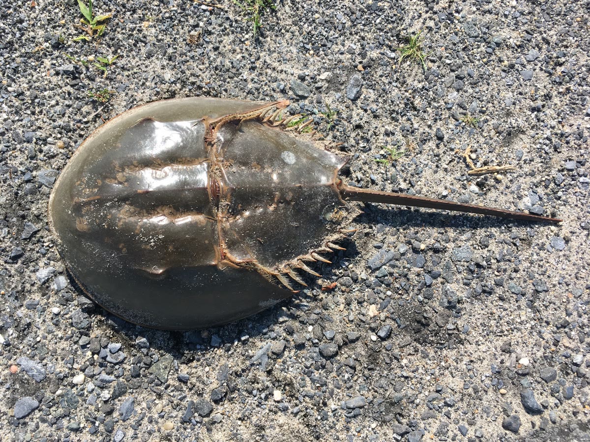
Our blood is red because of iron; the blood of a horseshoe crab is colourless in its veins and turns blue in air, because it carries oxygen on copper. Life uses a handful of metal ions for the jobs no organic group can do: binding oxygen reversibly, moving single electrons, splitting water, reducing nitrogen, making a water molecule acidic enough to attack carbon dioxide. This chapter reads those jobs with the tools of coordination chemistry: hard and soft acids, ligand fields and spin states, equilibria and rate laws, and it ends with the metals used as drugs.
You already know
The Year 2 volume treated -amino acids, side chains and peptides, nucleobases and nucleotides, coordination complexes, ligand fields, high and low spin, and hard and soft nucleophiles and electrophiles; the Year 1 volume complexation constants and the pL scale. Chapter 18 and Chapter 19 gave spectra, Marcus theory and the aquation of cisplatin, Chapter 13 Michaelis–Menten kinetics, and Chapter 8 the relaxation time .
24.1 Metals in biology
Definition 24.1 (Metalloproteins)
A metalloprotein is a protein that contains bound metal ions; a metalloenzyme is one that catalyses a reaction. A cofactor is a non-protein species, a metal ion or a small molecule, bound to a protein and needed for its activity.
The metals that matter in quantity are sodium, potassium, magnesium and calcium, which mostly carry charge and hold structures; the transition metals iron, zinc, copper, manganese, cobalt, molybdenum and nickel occur in traces but run much of the chemistry. A protein chooses its metal through the donor atoms it offers and their geometry.
Definition 24.2 (Hard and soft acids)
A hard acid is a small, highly charged, weakly polarisable Lewis acid (, , , ); a soft acid is a large, polarisable one of low charge (, , , ). The hard and soft acid–base principle states that hard acids bind preferably to hard bases (oxygen donors, fluoride) and soft acids to soft bases (sulfur donors, iodide).
In proteins, carboxylates and phenolates are hard, the thiolate of cysteine and the thioether of methionine soft, and the imidazole of histidine in between. Iron(III) sits among oxygen donors, copper(I) among sulfur donors, zinc, a borderline acid, with histidine and cysteine; mercury and cadmium are toxic in part because they seize the cysteines of enzymes.
Definition 24.3 (Irving–Williams series)
The Irving–Williams series is the order of stability of the high-spin complexes of the divalent ions of the first transition series with a given ligand: .
Proposition 24.4 (Irving–Williams order)
The series holds for almost all ligands (an experimental observation); it follows the decrease of ionic radius from manganese to copper, the ligand-field stabilisation energy, zero for , largest for , and the Jahn–Teller distortion of copper(II), which shortens four of its bonds.
Argued. The radius falls across the row as the nuclear charge grows, which strengthens the electrostatic attraction of every ligand. The ligand-field stabilisation of high-spin octahedral ions (Chapter 18) is , , , and for to , and for : it adds to the trend up to nickel and drops for zinc; copper(II) recovers more than its stabilisation suggests by its tetragonal distortion, which gives four short, strong bonds. ∎
A consequence that cells must manage: copper and zinc would outcompete the other ions for most sites, so their free concentrations are kept extremely low by dedicated carrier proteins.
24.2 Oxygen transport
Definition 24.5 (Porphyrins and haem)
A porphyrin is a large, flat, aromatic macrocycle of four pyrrole rings joined by methine bridges, whose four nitrogens bind a metal ion at its centre. Haem is the iron complex of protoporphyrin IX, the cofactor of haemoglobin, myoglobin and the cytochromes.
In myoglobin and haemoglobin, the iron of haem has a fifth ligand below the ring, the nitrogen of the proximal histidine, and a sixth site free for . Deoxy iron(II) is high spin: an electron in the orbital, pointing at the four nitrogens, makes it too large for the hole of the ring, and it sits out of the plane, towards the histidine. On binding it becomes low spin, smaller, and moves into the plane, pulling the histidine and the protein helix with it.
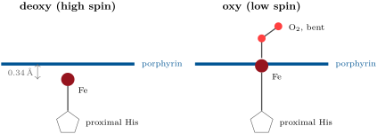
Bound dioxygen is better described as superoxide on iron(III), , than as neutral on iron(II): the complex is diamagnetic because the unpaired electrons of the two centres couple. The protein stops the irreversible oxidation to iron(III) and water, which a bare haem in water undergoes at once.
Definition 24.6 (Cooperative binding)
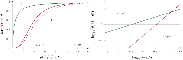
Binding to a protein with several sites is cooperative when the binding of one ligand raises the affinity of the remaining sites. The Hill coefficient is the slope of the Hill plot, against , at half saturation ( is the fraction of sites occupied).
Proposition 24.7 (One site)
For a protein with one site, , the fraction occupied is , where is the dissociation constant expressed as a pressure.
Proof. , so , and . ∎
Theorem 24.8 (Hill equation)
For a protein with sites that are either all empty or all filled, ,
Proof. ; write . The fraction of sites occupied is that of molecules in the filled form, . Then , and taking logarithms gives the straight line of slope . ∎
Haemoglobin, with four sites, is not fully cooperative: its Hill coefficient is well below 4, about 3, and its Hill plot has slope 1 at both ends, where it behaves as a deoxy or as an oxy protein with independent sites. The cooperativity comes from a change of quaternary structure: the motion of the iron into the plane of the first haem to bind is passed to the other subunits.
Method 24.9 (Reading a Hill plot)
- From each measured saturation compute ; plot it against .
- The pressure where the plot crosses zero is .
- The slope at that point is the Hill coefficient: means independent sites, cooperative binding.
- With two points only, .
Haemoglobin also binds protons and carbon dioxide at sites away from the haem, which shift its curve to the right where they are abundant, in working tissues: the Bohr effect releases more oxygen exactly where it is needed.
Proposition 24.10 (Carbon monoxide competition)
If carbon monoxide and dioxygen compete for the same sites, with of equilibrium constant , then .
Proof. At equilibrium ; rearranging gives the result. ∎
is in the hundreds for human haemoglobin, so that a few hundred parts per million of carbon monoxide occupy a large fraction of the sites; worse, the sites left free hold their oxygen more tightly, as the cooperativity works on them. Free haem binds carbon monoxide far more strongly still: in the protein, a distal histidine above the iron hinders the linear Fe–C–O geometry carbon monoxide prefers, while it hydrogen-bonds the bent . Other animals use other metals: the haemocyanins of molluscs and arthropods bind between two copper ions as peroxide, the haemerythrins of some marine worms on a pair of iron ions.
In the lab — An oxygen-binding curve
A solution of haemoglobin in buffer is placed in a gas-tight cuvette fitted with a side flask. It is first freed of oxygen by flushing with nitrogen, then known volumes of air are injected and the solution equilibrated by gentle tilting. After each addition the visible spectrum is recorded: the bands of the deoxy form (one band near ) give way to the two bands of the oxy form, and the saturation is read from the absorbance at one wavelength, between its deoxy and oxy limits.
24.3 Metalloenzymes
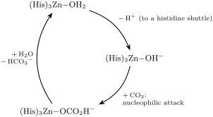
Carbonic anhydrase speeds up , which in water alone takes seconds, enough to matter in the red cell’s short passage through a lung capillary. Its zinc ion is held by three histidines and a water molecule. A small cation of high charge, it lowers the of that water from about 14 to about 7: at physiological pH a good part of the enzyme carries a zinc-bound hydroxide, a strong nucleophile, held right next to a pocket that binds .
Proposition 24.11 (A fast enzyme)
The specificity constant of carbonic anhydrase approaches the rate constant of encounter of with the enzyme: nearly every encounter leads to reaction.
Argued. is the second-order rate constant of enzyme and substrate at low substrate concentration (Chapter 13); it cannot exceed the diffusion-controlled rate constant of their encounter (Chapter 12). The measured values for carbonic anhydrase are among the highest known for any enzyme and approach that limit: the chemistry inside the active site is then no longer what limits the rate. ∎
Cytochrome P450 enzymes, in the liver and in many other cells, hydroxylate C–H bonds of drugs and hormones. Their haem iron is held by a cysteine thiolate; with and two electrons it forms an iron(IV)-oxo complex with a radical on the porphyrin, called compound I, which abstracts a hydrogen atom from the substrate and returns the hydroxyl group to the carbon radical. Nitrogenase reduces to ammonia at room temperature on a cluster of seven iron atoms, one molybdenum and nine sulfides around a central carbon atom, the FeMo cofactor, at the cost of many molecules of ATP. Vitamin contains a cobalt–carbon bond, one of the very few metal–carbon bonds in biology, whose homolysis starts radical rearrangements.
24.4 Electron transfer and energy
Proteins that only move electrons have metal centres with small reorganisation energies (Chapter 19): the two oxidation states have almost the same geometry, so that the Marcus barrier is low.
Definition 24.12 (Iron–sulfur clusters)
An iron–sulfur cluster is a group of iron ions bridged by sulfide ions and bound to the protein by cysteine thiolates: , and the cubane-like .
Definition 24.13 (Blue copper proteins)
A blue copper protein carries one copper ion bound by two histidines, a cysteine and a weakly bound methionine in a distorted tetrahedral site; its intense blue colour comes from a cysteine-to-copper(II) charge-transfer band.
Definition 24.14 (Entatic state)
The entatic state is a metal site held by the protein in a strained geometry, between the geometries preferred by its two oxidation states, which lowers the reorganisation energy of its reactions.
Copper(II) alone prefers a square plane, copper(I) a tetrahedron: in the blue copper site neither is satisfied, and electron transfer is fast.
Definition 24.15 (Oxygen-evolving complex)
The oxygen-evolving complex of photosystem II is the cluster that oxidises water to dioxygen in photosynthesis.
Proposition 24.16 (Four photons per oxygen)
Oxidising two water molecules to one requires four photons absorbed by photosystem II, the cluster passing through five states to .
Proof. : four electrons must be removed. Each photon absorbed by the reaction centre moves one electron away from it, and the cluster refills the oxidised centre one electron at a time: four photons, four steps, storing oxidising equivalents in the states to ; releases and returns to . ∎
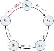
At the other end of the energy chain, cytochrome oxidase of the mitochondria reduces to water with four electrons and four protons on a haem iron and a copper ion facing each other, without releasing the partly reduced, damaging superoxide or peroxide.
24.5 Metals in medicine
Definition 24.17 (Metallodrugs)
A metallodrug is a drug whose activity depends on a metal ion or a metal complex.
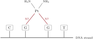
Cisplatin, cis-, is stable in blood, where the chloride concentration is high; inside cells, where it is much lower, it exchanges a chloride for water (Chapter 19). The aqua complex binds the N7 atom of guanine, then a second guanine next to it on the same strand: the 1,2-GG cross-link bends the DNA, which the cell cannot repair, and it dies. The trans isomer cannot bridge two neighbouring bases and is inactive. Carboplatin, with a chelating dicarboxylate that leaves slowly, has fewer side effects; oxaliplatin, with a diaminocyclohexane ligand, works on tumours resistant to cisplatin.
Definition 24.18 (Contrast agents)
A contrast agent for magnetic resonance imaging is a paramagnetic compound that shortens the relaxation times of nearby water protons. Its relaxivity is the increase of the relaxation rate per unit concentration of the agent.
Proposition 24.19 (Relaxation rate)
With an agent of relaxivity at concentration , the relaxation rate of water protons is , where is the value without agent.
Proof. The paramagnetic contribution is proportional to the number of agent molecules that water molecules visit per unit time, hence to ; relaxation rates from independent mechanisms add. By definition of the added rate is . ∎
Gadolinium(III), with seven unpaired electrons and a slowly relaxing electron spin, is the metal of choice. Free is toxic: it is given as a very stable complex of an octadentate polyaminocarboxylate that leaves one site for a water molecule, which exchanges fast with the bulk and carries the relaxation to all of it.
Definition 24.20 (Chelation therapy)
Chelation therapy is the removal of a toxic metal from the body by a chelating ligand given as a drug, which forms a stable, soluble complex that is excreted.
Method 24.21 (Choosing a chelating drug)
- Compare the stability constants of the chelator with the toxic metal and with the essential metals present in large amounts (, , ).
- Compute the free metal level the chelator leaves at the concentrations and pH of the body: the higher pM for the toxic metal and the lower for the essential ones, the better.
- Give the chelator already loaded with the competing essential ion (the calcium complex of EDTA for lead), so that it exchanges rather than strips calcium from the blood.
- Check the hard–soft match (sulfur donors for mercury and lead, hard oxygen donors for iron(III)) and that the complex is excreted.
Safety


Cisplatin: fatal if swallowed, may cause cancer and genetic defects, may damage fertility, causes serious eye damage and allergic reactions; it is handled only as a prepared drug solution, with gloves, in a ventilated cabinet.
History — A protein structure and an accidental drug
Max Perutz worked for more than twenty years on the X-ray structure of haemoglobin, solved at low resolution in 1959; with John Kendrew, who solved myoglobin, he received the 1962 Nobel Prize in Chemistry. In 1965 Barnett Rosenberg, passing a current between platinum electrodes in a culture of bacteria, saw them grow into long filaments without dividing; the cause was not the current but a platinum ammine complex formed from the electrodes. It became cisplatin, still one of the most used anticancer drugs.
24.6 Exercises
Exercise 24.1 ★
A protein is 20 % saturated at of oxygen and 80 % at . Compute its Hill coefficient.
Solution
Solution of Exercise 24.1.
goes from to while goes up by : .
Exercise 24.2 ★
Order , , , , and by the stability of their complexes with a given ligand, and explain the place of zinc.
Solution
Solution of Exercise 24.2.
. Zinc () has no ligand-field stabilisation and no Jahn–Teller distortion: it falls back below copper.
Exercise 24.3 ★
Predict the preferred protein donors (carboxylate, cysteine thiolate, histidine imidazole) of , , , and .
Solution
Solution of Exercise 24.3.
and , hard: carboxylates (and phenolates for iron). and , soft: cysteine thiolates. , borderline: histidine and cysteine.
Exercise 24.4 ★
How many electrons are removed, and how many photons absorbed by photosystem II, per molecule of evolved? Per mole?
Solution
Solution of Exercise 24.4.
: four electrons, four photons per ; four moles of photons per mole of .
Exercise 24.5 ★★
With the model curves of the chapter ( for myoglobin; and for haemoglobin), compute the fraction of its oxygen each protein gives up between (lungs) and (tissues).
Solution
Solution of Exercise 24.5.
Myoglobin: and : it gives up % of its oxygen. Haemoglobin: and : it gives up 26 %.
Exercise 24.6 ★★
Air containing of carbon monoxide is breathed for long enough to reach equilibrium. With (data of the exercise) and that of the air (20.95 % oxygen), what fraction of the haemoglobin carries CO?
Solution
Solution of Exercise 24.6.
; fraction %.
Exercise 24.7 ★★
Give the formal oxidation states of the iron ions in the clusters , and (SR: cysteinate, S: sulfide).
Solution
Solution of Exercise 24.7.
Sulfides each, cysteinates each. : irons sum : two Fe(III). : , one Fe(III) and one Fe(II) (mixed valence). : , two Fe(III) and two Fe(II), each iron on average.
Exercise 24.8 ★★
Write the first aquation of cisplatin, and explain why it is slow in blood and faster inside cells. Which atom of DNA does platinum bind, and why is the trans isomer inactive?
Solution
Solution of Exercise 24.8.
. The high chloride concentration of blood pushes the equilibrium back; inside cells chloride is much lower, and the aqua complex forms. Platinum binds N7 of guanine. In the trans isomer the two labile sites are opposite each other and cannot reach two adjacent bases on one strand.
Exercise 24.9 ★★
A tissue has . A gadolinium agent of relaxivity reaches in it (data of the exercise). Compute the new .
Solution
Solution of Exercise 24.9.
: .
Exercise 24.10 ★★★
A chelator L has with and with (data of the exercise). Compute the equilibrium constant of and explain why the calcium complex is given rather than the free ligand.
Solution
Solution of Exercise 24.10.
: lead displaces calcium completely. The free ligand would also bind the calcium of the blood and lower its concentration dangerously; the calcium complex exchanges with lead and leaves the calcium level unchanged.
Exercise 24.11 ★★★
Carbonic anhydrase has (data of the exercise), a typical enzyme about . At , far below , compare the time each needs to convert half the substrate with an enzyme concentration of .
Solution
Solution of Exercise 24.11.
Far below the reaction is first order: , . Carbonic anhydrase: , . Typical enzyme: , , a thousand times longer, too slow for the second a red cell spends in a lung capillary.
Exercise 24.12 ★★★
With , what pressure of carbon monoxide occupies half the sites in the presence of air at of oxygen? Express it in ppm of a gas at .
Solution
Solution of Exercise 24.12.
Half the sites with CO means : , about .
24.7 Problem: How Much Oxygen Does Blood Deliver?
Problem 24.1
Weekend problem — how much oxygen does blood deliver? Hill curves of haemoglobin and myoglobin, the oxygen carried by a litre of blood, the Bohr shift, and what carbon monoxide takes away
Model data of the problem: haemoglobin and at pH 7.4, at the lower pH of working tissues; myoglobin ; blood contains of haemoglobin, of molar mass , with four sites per molecule; is in the lungs and in the tissues; the heart pumps at rest; for carbon monoxide. Gas volumes at and .
Part I — The curves.
- Compute the saturation of haemoglobin in the lungs.
- Compute it in the tissues at pH 7.4.
- Compute the saturation of myoglobin at .
- Why can myoglobin take oxygen from haemoglobin in a muscle?
- What Hill coefficient would four independent sites give?
- Explain in one sentence where the cooperativity comes from.
Part II — A litre of blood.
- Compute the concentration of haemoglobin in mol/L.
- Compute the concentration of oxygen sites.
- Compute the oxygen carried per litre in the lungs, in mmol.
- Compute the oxygen delivered per litre to the tissues at pH 7.4.
- Convert it to millilitres of gas.
- What fraction of the oxygen carried is that?
Part III — The Bohr shift.
- Compute the saturation in the tissues at the lower pH.
- Compute the oxygen delivered per litre with the shift.
- By what factor does the shift increase delivery?
- What lowers the pH of a working muscle?
- Compute the oxygen delivered per minute at rest, in mmol.
- Convert it to litres of gas per minute.
Part IV — Carbon monoxide.
- Compute for air with of CO.
- Compute the fraction of sites lost to CO.
- Why is the loss of delivered oxygen worse than that fraction?
- Why is pure oxygen the first treatment?
- Why does the distal histidine matter for survival?
- State the result: the litres of oxygen delivered per minute at rest.
Solution
Solution of Problem 24.1.
1. .
2. .
3. .
4. Its is ten times lower: at the pressures of a working muscle it is far more saturated than haemoglobin at the same pressure, so oxygen passes from haemoglobin to myoglobin.
5. .
6. Binding at one haem moves its iron into the plane and pulls the histidine and its helix; the subunits shift together into a form of higher affinity for the other sites.
7. .
8. .
9. .
10. .
11. .
12. %.
13. .
14. .
15. : thirty per cent more.
16. Carbon dioxide made by respiration, hydrated to carbonic acid by carbonic anhydrase, and lactic acid in hard work.
17. .
18. .
19. .
20. : about 10 % of the sites.
21. The sites still carrying oxygen, in a molecule that also carries CO, are in the high-affinity form: the curve shifts to the left and they release less of their oxygen in the tissues.
22. By the competition relation the ratio falls in proportion to : breathing pure oxygen, almost five times the pressure in air, displaces carbon monoxide faster.
23. By hindering the linear binding of CO and hydrogen-bonding , it keeps in the hundreds instead of the much larger value of free haem; otherwise even the carbon monoxide made in the body would block much of the haemoglobin.
24. At rest the blood delivers about of oxygen per minute.